
CRITICAL CARE - ADULTS / SPECIAL ARTICLE
Central venous cannulation in critically ill patients: guidelines of the Polish Society of Anaesthesiology and Intensive Therapy
1
2nd Department of Anaesthesiology and Intensive Care, Medical University of Warsaw, Poland
2
Department of Anesthesiology and Intensive Care, Institute of Medical Sciences, University of Opole, Poland
3
Clinical Department of Gynecology, Gynecologic Oncology and Reproduction, National Medical Institute of the Ministry of the Interior and Administration, Warsaw, Poland
4
Department of Anaesthesiology and Intensive Care, Medical University of Gdansk, Poland
5
Department of Anaesthesiology and Intensive Care, Regional Hospital in Gorzow Wielkopolski, Poland
6
Department of Anaesthesiology and Intensive Care, Faculty of Medical Sciences in Zabrze, Medical University of Silesia
in Katowice, Zabrze, Poland
7
Faculty of Medicine and Health Sciences, Jan Kochanowski University, Kielce, Poland
8
Department of Anaesthesiology and Intensive Care Education, Medical University of Warsaw, Poland
9
Centre for Intensive Care and Perioperative Medicine, Jagiellonian University Medical College, Krakow, Poland
10
Clinic of Intensive Care and Anesthesiology, 5th Military Clinical Hospital with Polyclinic in Krakow, Poland
Submission date: 2026-03-03
Acceptance date: 2026-04-07
Publication date: 2026-05-21
Corresponding author
Mateusz Zawadka
2nd Department of Anaesthesiology and Intensive Care, Medical University of Warsaw, 1a Banacha St., 02-097 Warsaw, Poland
2nd Department of Anaesthesiology and Intensive Care, Medical University of Warsaw, 1a Banacha St., 02-097 Warsaw, Poland
Anaesthesiol Intensive Ther 2026;58(1):84-105
KEYWORDS
catheterizationcentral venousultrasonographycritical illnessintensive care unitspractice guidelines as topicvascular access devices
TOPICS
ABSTRACT
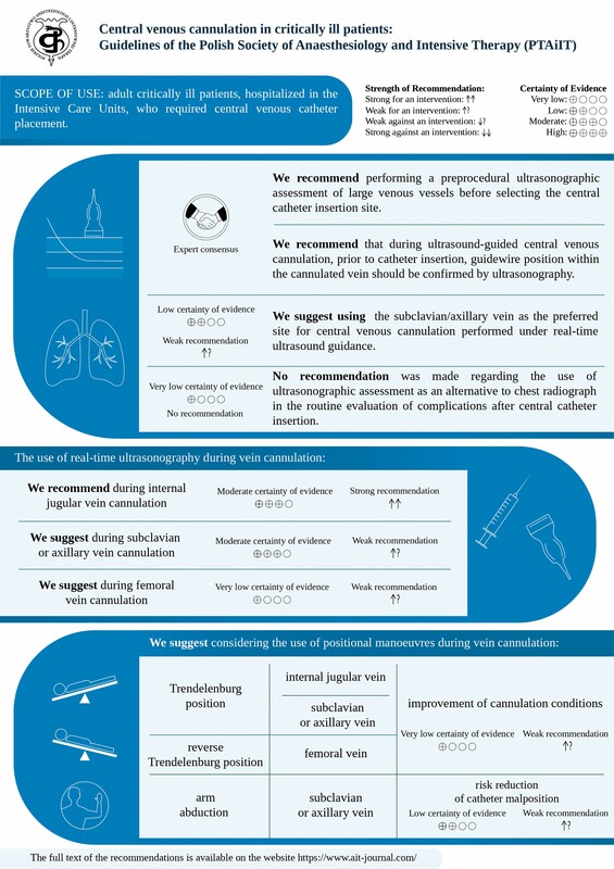
These guidelines provide evidence-based recommendations for central venous cannulation in critically ill patients in the intensive care unit. The document was developed by the Working Group of the Polish Society of Anaesthesiology and Intensive Therapy (Polskie Towarzystwo Anestezjologii i Intensywnej Terapii – PTAiIT) based on the GRADE (Grading of Recommendations Assessment, Development and Evaluation) methodology, which encompasses systematic reviews of the literature, meta-analyses, and – in the absence of sufficient data – expert consensus. These guidelines aim to standardise the approach to central venous cannulation, increase the effectiveness of procedures, and minimise the risk of complications. The guidelines address pre-procedural ultrasound assessment, selection of the optimal vascular access site, comparison of ultrasound-guided versus landmark cannulation at different access sites, confirmation of catheter position after cannulation, and the role of positional manoeuvres.
ADDITIONAL INFORMATION
Polish version available HERE
REFERENCES (101)
1.
Guyatt GH, Oxman AD, Vist GE, Kunz R, Falck-Ytter Y, Alonso-Coello P, Schünemann HJ; for the GRADE Working Group. GRADE: an emerging consensus on rating certainty of evidence and strength of recommendations. BMJ 2008; 336: 924-926. DOI: 10.1136/bmj.39489.470347.AD.
2.
Teja B, Bosch NA, Diep C, Pereira TV, Mauricio P, Sklar MC, et al. Complication rates of central venous catheters: a systematic review and meta-analysis. JAMA Intern Med 2024; 184: 474-482. DOI: 10.1001/jamainternmed.2023.8232.
3.
Zawadka M, Andruszkiewicz P, Gola W, Wong A, Czuczwar M. Echocardiography and Ultrasound Committee statement for the accreditation programme in point-of-care ultrasonography in Poland. Anaesthesiol Intensive Ther 2023; 55: 77-80. DOI: 10.5114/ait.2023.128704.
4.
Robba C, Wong A, Poole D, Al Tayar A, Arntfield RT, Chew MS, et al. Basic ultrasound head-to-toe skills for intensivists in the general and neuro intensive care unit population: consensus and expert recommendations of the European Society of Intensive Care Medicine. Intensive Care Med 2021; 47: 1347-1367. DOI: 10.1007/s00134-021-06486-z.
5.
Annetta MG, Spencer TR, Pittiruti M. The never-ending confusion between subclavian vein and axillary vein. J Vasc Access 2025. DOI: 10.1177/11297298251352477.
6.
Gawda R, Czarnik T. Ultrasound-guided infraclavicular cannulation of the subclavian vein – still an ongoing misconception. J Intensive Care Soc 2023; 24 (3 Suppl): 10. DOI: 10.1177/1751143720914188.
7.
Sterne JAC, Savović J, Page MJ, Elbers RG, Blencowe NS, Boutron I, et al. RoB 2: a revised tool for assessing risk of bias in randomised trials. BMJ 2019; 366: l4898. DOI: 10.1136/bmj.l4898.
8.
Whiting PF, Rutjes AWS, Westwood ME, Mallett S, Deeks JJ, Reitsma JB, et al. QUADAS-2: a revised tool for the quality assessment of diagnostic accuracy studies. Ann Intern Med 2011; 155: 529-536. DOI: 10.7326/0003-4819-155-8-201110180-00009.
9.
Nasa P, Jain R, Juneja D. Delphi methodology in healthcare research: how to decide its appropriateness. World J Methodol 2021; 11: 116-129. DOI: 10.5662/wjm.v11.i4.116.
10.
Sakuraya M, Okano H, Yoshihiro S, Niida S, Kimura K. Insertion site of central venous catheter among hospitalized adult patients: a systematic review and network meta-analysis. Front Med (Lausanne) 2022; 9: 960135. DOI: 10.3389/fmed.2022.960135.
11.
Merrer J. Complications of femoral and subclavian venous catheterization in critically ill patients. A randomized controlled trial. JAMA 2001; 286: 700. DOI: 10.1001/jama.286.6.700.
12.
Gülmen Ş, Kiriş Ý, Peker O, Koçyiğit A, Okutan H, Kuralay E, Ocal A. Central venous catheterization in open heart surgery: internal jugular vein or supraclavicular subclavian vein approach? Turk J Thorac Cardiovasc Surg 2010; 18: 11-16.
13.
Kocum A, Sener M, Calıskan E, Bozdogan N, Atalay H, Aribogan A. An alternative central venous route for cardiac surgery: supraclavicular subclavian vein catheterization. J Cardiothorac Vasc Anesth 2011; 25: 1018-1023. DOI: 10.1053/j.jvca.2011.02.006.
14.
Parienti JJ, Mongardon N, Mégarbane B, Mira JP, Kalfon P, Gros A, et al. Intravascular complications of central venous catheterization by insertion site. N Engl J Med 2015; 373: 1220-1229. DOI: 10.1056/NEJMoa1500964.
15.
Laiq N, Majid A, Nawab J, Malik A. Central venous catheterization and cardiac surgeries. J Med Sci 2015; 23: 137-140.
16.
Shin HJ, Na HS, Koh WU, Ro YJ, Lee JM, Choi YJ, et al. Complications in internal jugular vs subclavian ultrasound-guided central venous catheterization: a comparative randomized trial. Intensive Care Med 2019; 45: 968-976. DOI: 10.1007/s00134-019-05651-9.
17.
Fournil C, Boulet N, Bastide S, Louart B, Ambert A, Boutin C, et al. High success rates of ultrasound‐guided distal internal jugular vein and axillary vein approaches for central venous catheterization: a randomized controlled open‐label pilot trial. J Clin Ultrasound 2023; 51: 158-166. DOI: 10.1002/jcu.23383.
18.
Trabelsi B, Hajjej Z, Drira D, et al. Comparison of ultrasound-guided internal jugular vein and supraclavicular subclavian vein catheterization in critically ill patients: a prospective, randomized clinical trial. Ann Intensive Care 2022; 12: 91. DOI: 10.1186/s13613-022-01065-x.
19.
Czarnik T, Czuczwar M, Borys M, Chrzan O, Filipiak K, Maj M, et al. Ultrasound-guided infraclavicular axillary vein versus internal jugular vein cannulation in critically ill mechanically ventilated patients: a randomized trial. Crit Care Med 2023; 51: e37-e44. DOI: 10.1097/CCM.0000000000005740.
20.
Corradi F, Cucciolini G, Tavazzi G, Wong A, Balan C, Melniker LA, et al. WINFOCUS worldwide survey on central venous catheter insertion and position confirmation practices (CVC-ICON study). Ultrasound J 2025; 17: 41. DOI: 10.1186/s13089-025-00429-1.
21.
Brass P, Hellmich M, Kolodziej L, Schick G, Smith AF. Ultrasound guidance versus anatomical landmarks for internal jugular vein catheterization. Cochrane Database Syst Rev 2015; 1: CD006962. DOI: 10.1002/14651858.CD006962.pub2.
22.
Faiz S, Rahimzadeh P, Ziyaeifard M, Hassani V. Comparison between ultrasound guidance and the landmark technique for the internal jugular vein cannulation in adult patients by anesthesia residents or inexperienced operators. Iranian Heart Journal 2018; 19: 30-37.
23.
Singh AP, Singh DK, Agarwal A. Ultrasonography: a novel approach to central venous cannulation. Indian J Crit Care Med 2009; 13: 213-216. DOI: 10.4103/0972-5229.60174.
24.
Bansal R, Agarwal SK, Tiwari SC, Dash SC. A prospective randomized study to compare ultrasound-guided with nonultrasound-guided double lumen internal jugular catheter insertion as a temporary hemodialysis access. Ren Fail 2005; 27: 561-564. DOI: 10.1080/08860220500199084.
25.
Dolu H, Goksu S, Sahin L, Ozen O, Eken L. Comparison of an ultrasound-guided technique versus a landmark-guided technique for internal jugular vein cannulation. J Clin Monit Comput 2015; 29: 177-182. DOI: 10.1007/s10877-014-9585-3.
26.
Fathi M, Izanloo A, Jahanbakhsh S, Gilani MT, Majidzadeh A, Benhangi AS, Paravi N, et al. Central venous cannulation of the internal jugular vein using ultrasound-guided and anatomical landmark techniques. Anesth Pain Med 2016; 6: e35803. DOI: 10.5812/aapm.35803.
27.
Saiprabha S, Mohamed Sameer B, Sathish Madhavan T. Comparative study of right internal jugular vein cannulation by the real-time ultrasound imaging, ultrasound-guided prelocation and the anatomical landmark technique – a prospective randomized trial. Int J Acad Med Pharm 2023; 5: 750-754. DOI: 10.47009/jamp.2023. 5.4.149.
28.
Nazari I, Musavi M, Alavi M. Comparing outcomes and complication of central venous cannulation using both conventional and ultrasound guide. Biosci Biotechnol Res Asia 2015; 12: 2167-2171. DOI: 10.13005/bbra/1888.
29.
Ehtesham AM, Patkar C, Phalgune D. Study between ultrasound guided technique and conventional landmark technique for internal jugular vein cannulation: a randomised controlled trial. J Clin Diagn Res 2020; 13: 9-12. DOI: 10.7860/JCDR/2020/43578.13597.
30.
Ahmed SS, Samad K, Yousuf MS, Qamar-ul-Hoda M. A comparison of techniques of internal jugular vein cannulation: anatomical landmark, ultrasound guided pre-location, and real-time ultrasound guided. Cureus 2024; 16: e54499. DOI: 10.7759/cureus.54499.
31.
Hayashi H, Tsuzuku M, Amano M. Simplfied echo‐guided internal jugular vein puncture: a comparison to the landmark‐guided technique. Anesth Analg 1998; 86. DOI: 10.1097/00000539-199804001-00089.
32.
Hayashi H, Amano M. Does ultrasound imaging before puncture facilitate internal jugular vein cannulation? Prospective randomized comparison with landmark-guided puncture in ventilated patients. J Cardiothorac Vasc Anesth 2002; 16: 572-575. DOI: 10.1053/jcan.2002.126950.
33.
Karakitsos D, Labropoulos N, De Groot E, Patrianakos AP, Kouraklis G, Poularas J, et al. Real-time ultrasound-guided catheterisation of the internal jugular vein: a prospective comparison with the landmark technique in critical care patients. Crit Care 2006; 10: R162. DOI: 10.1186/cc5101.
34.
Leung J, Duffy M, Finckh A. Real-time ultrasonographically-guided internal jugular vein catheterization in the emergency department increases success rates and reduces complications: a randomized, prospective study. Ann Emerg Med 2006; 48: 540-547. DOI: 10.1016/ j.annemergmed.2006.01.011.
35.
Milling TJ, Rose J, Briggs WM, Birkhahn R, Gaeta TJ, Bove JJ, Melniker LA. Randomized, controlled clinical trial of point-of-care limited ultrasonography assistance of central venous cannulation: the Third Sonography Outcomes Assessment Program (SOAP-3) Trial. Crit Care Med 2005; 33: 1764-1769. DOI: 10.1097/01.ccm. 0000171533.92856.e5.
36.
Palepu GB, Deven J, Subrahmanyam M, Mohan S. Impact of ultrasonography on central venous catheter insertion in intensive care. Indian J Radiol Imaging 2009; 19: 191-198. DOI: 10.4103/0971-3026.54877.
37.
Soyer P, Lacheheb D, Levesque M. High-resolution sonographic guidance for transjugular liver biopsy. Abdom Imaging 1993; 18: 360-362. DOI: 10.1007/BF00201782.
38.
Sulek CA, Blas ML, Lobato EB. A randomized study of left versus right internal jugular vein cannulation in adults. J Clin Anesth 2000; 12: 142-145. DOI: 10.1016/s0952-8180(00)00129-x.
39.
Teichgräber UK, Benter T, Gebel M, Manns MP. A sonographically guided technique for central venous access. AJR Am J Roentgenol 1997; 169: 731-733. DOI: 10.2214/ajr.169.3.9275887.
40.
Troianos CA, Jobes DR, Ellison N. Ultrasound guided cannulation of the internal jugular vein. Anesthesiology 1990; 73 (3A): A451.
41.
Troianos CA, Jobes DR, Ellison N. Ultrasound-guided cannulation of the internal jugular vein. A prospective, randomized study. Anesth Analg 1991; 72: 823-826. DOI: 10.1213/00000539-199106000-00020.
42.
Turker G, Kaya FN, Gurbet A, Aksu H, Erdogan C, Atlas A. Internal jugular vein cannulation: an ultrasound-guided technique versus a landmark-guided technique. Clinics 2009; 64: 989-992. DOI: 10.1590/S1807-59322009001000009.
43.
Mallory DL, McGee WT, Shawker TH, Brenner M, Bailey KR, Evans RG, et al. Ultrasound guidance improves the success rate of internal jugular vein cannulation. A prospective, randomized trial. Chest 1990; 98: 157-160. DOI: 10.1378/chest.98.1.157.
44.
Kornbau C, Lee KC, Hughes GD, Firstenberg MS. Central line complications. Int J Crit Illn Inj Sci 2015; 5: 170-178. DOI: 10.4103/2229-5151.164940.
45.
Zawadka M, La Via L, Wong A, Olusanya O, Muscarà L, Continella C, et al. Real-time ultrasound guidance as compared with landmark technique for subclavian central venous cannulation: a systematic review and meta-analysis with trial sequential analysis. Crit Care Med 2023; 51: 642-652. DOI: 10.1097/CCM.0000000000005819.
46.
Singam AP, Chaudhary A, Shrey S. Anatomical landmark guided versus ultrasound-guided technique for subcla- vian vein cannulation in critically ill patients. JKIMSU 2019; 8: 50-57.
47.
Gualtieri E, Deppe SA, Sipperly ME, Thompson DR. Subclavian venous catheterization: greater success rate for less experienced operators using ultrasound guidance. Crit Care Med 1995; 23: 692-697. DOI: 10.1097/00003246-199504000-00018.
48.
Fragou M, Gravvanis A, Dimitriou V, Papalois A, Kouraklis G, Karabinis A, et al. Real-time ultrasound-guided subclavian vein cannulation versus the landmark method in critical care patients: a prospective randomized study. Crit Care Med 2011; 39: 1607-1612. DOI: 10.1097/CCM.0b013e318218a1ae.
49.
Sazdov D, Srceva MJ, Todorova ZN. Comparative analysis of ultrasound guided central venous catheterization compared to blind catheterization. Pril (Makedon Akad Nauk Umet Odd Med Nauki) 2017; 38: 107-114. DOI: 10.1515/prilozi-2017-0028.
50.
Subramony R, Spann R, Medak A, Campbell C. Ultrasound-guided vs. landmark method for subclavian vein catheterization in an academic emergency department. J Emerg Med 2022; 62: 760-768. DOI: 10.1016/j.jemermed.2021.11.002.
51.
Su Y, Hou JY, Ma GG, et al. Comparison of the proximal and distal approaches for axillary vein catheterization under ultrasound guidance (PANDA) in cardiac surgery patients susceptible to bleeding: a randomized controlled trial. Ann Intensive Care 2020; 10: 90. DOI: 10.1186/s13613-020-00703-6.
52.
Brass P, Hellmich M, Kolodziej L, Schick G, Smith AF. Ultrasound guidance versus anatomical landmarks for subclavian or femoral vein catheterization. Cochrane Database Syst Rev 2015; 1: CD011447. DOI: 10.1002/14651858.CD011447.
53.
Prabhu M V, Juneja D, Gopal PB, Sathyanarayanan M, Subhramanyam S, Gandhe S, Nayak KS. Ultrasound-guided femoral dialysis access placement: a single-center randomized trial. Clin J Am Soc Nephrol 2010; 5: 235-239. DOI: 10.2215/CJN.04920709.
54.
Smit JM, Raadsen R, Blans MJ, Petjak M, Van de Ven PM, Tuinman PR. Bedside ultrasound to detect central venous catheter misplacement and associated iatrogenic complications: a systematic review and meta- analysis. Crit Care 2018; 22: 65. DOI: 10.1186/s13054-018-1989-x.
55.
Blans MJ, Endeman H, Bosch FH. The use of ultrasound during and after central venous catheter insertion versus conventional chest X-ray after insertion of a central venous catheter. Neth J Med 2016; 74: 353-357.
56.
Bedel J, Vallée F, Mari A, Riu B, Planquette B, Geeraerts T, et al. Guidewire localization by transthoracic echocardiography during central venous catheter insertion: a periprocedural method to evaluate catheter placement. Intensive Care Med 2013; 39: 1932-1937. DOI: 10.1007/s00134-013-3097-3.
57.
Kim SC, Gräff I, Sommer A, Hoeft A, Weber S. Ultrasound-guided supraclavicular central venous catheter tip positioning via the right subclavian vein using a microconvex probe. J Vasc Access 2016; 17: 435-439. DOI: 10.5301/jva.5000518.
58.
Kim SC, Heinze I, Schmiedel A, Baumgarten G, Knuefermann P, Hoeft A, Weber S. Ultrasound confirmation of central venous catheter position via a right supraclavicular fossa view using a microconvex probe: an observational pilot study. Eur J Anaesthesiol 2015; 32: 29-36. doi: 10.1097/EJA.0000000000000042.
59.
Meggiolaro M, Scatto A, Zorzi A, Roman-Pognuz E, Lauro A, Passarella C, Bonaccorso G. Confirmation of correct central venous catheter position in the preoperative setting by echocardiographic “bubble-test”. Minerva Anestesiol 2015; 81: 989-1000.
60.
Vezzani A, Brusasco C, Palermo S, Launo C, Mergoni M, Corradi F. Ultrasound localization of central vein catheter and detection of postprocedural pneumothorax: an alternative to chest radiography. Crit Care Med 2010; 38: 533-538. DOI: 10.1097/CCM. 0b013e3181c0328f.
61.
Cortellaro F, Mellace L, Paglia S, Costantino G, Sher S, Coen D. Contrast enhanced ultrasound vs chest x-ray to determine correct central venous catheter position. Am J Emerg Med 2014; 32: 78-81. DOI: 10.1016/j.ajem.2013.10.001.
62.
Miccini M, Cassini D, Gregori M, Gazzanelli S, Cassibba S, Biacchi D. Ultrasound-guided placement of central venous port systems via the right internal jugular vein: are chest x-ray and/or fluoroscopy needed to confirm the correct placement of the device? World J Surg 2016; 40: 2353-2358. DOI: 10.1007/s00268-016-3574-2.
63.
Duran-Gehring PE, Guirgis FW, McKee KC, Goggans S, Tran H, Kalynych CJ, Wears RL. The bubble study: ultrasound confirmation of central venous catheter placement. Am J Emerg Med 2015; 33: 315-319. DOI: 10.1016/j.ajem.2014.10.010.
64.
Gekle R, Dubensky L, Haddad S, Bramante R, Cirilli A, Catlin T, et al. Saline flush test: can bedside sonography replace conventional radiography for confirmation of above-the-diaphragm central venous catheter placement? J Ultrasound Med 2015; 34: 1295-1299. DOI: 10.7863/ultra.34.7.1295.
65.
Kamalipour H, Ahmadi S, Kamali K, Moaref A, Shafa M, Kamalipour P. Ultrasound for localization of central venous catheter: a good alternative to chest X-ray? Anesth Pain Med 2016; 6: e38834. DOI: 10.5812/aapm.38834.
66.
Santarsia G, Casino FG, Gaudiano V, Mostacci SD, Bagnato G, Latorraca A, Lopez T. Jugular vein catheterization for hemodialysis: correct positioning control using real-time ultrasound guidance. J Vasc Access 2000; 1: 66-69. DOI: 10.1177/112972980000100207.
67.
Weekes AJ, Johnson DA, Keller SM, Efune B, Carey C, Rozario NL, Norton HJ, et al. Central vascular catheter placement evaluation using saline flush and bedside echocardiography. Acad Emerg Med 2014; 21: 65-72. DOI: 10.1111/acem.12283.
68.
Weekes AJ, Keller SM, Efune B, Ghali S, Runyon M. Prospective comparison of ultrasound and CXR for confirmation of central vascular catheter placement. Emerg Med J 2016; 33: 176-180. DOI: 10.1136/emermed-2015-205000.
69.
Wen M, Stock K, Heemann U, Aussieker M, Küchle C. Agitated saline bubble-enhanced transthoracic echocardiography: a novel method to visualize the position of central venous catheter. Crit Care Med 2014; 42: e231-e233. DOI: 10.1097/CCM.0000000000000130.
70.
Maury E, Guglielminotti J, Alzieu M, Guidet B, Offenstadt G. Ultrasonic examination: an alternative to chest radiography after central venous catheter insertion? Am J Respir Crit Care Med 2001; 164: 403-405. DOI: 10.1164/ajrccm.164.3.2009042.
71.
Zanobetti M, Coppa A, Bulletti F, Piazza S, Nazerian P, Conti A, et al. Verification of correct central venous catheter placement in the emergency department: comparison between ultrasonography and chest radiography. Intern Emerg Med 2013; 8: 173-180. DOI: 10.1007/s11739-012-0885-7.
72.
de Man L, Wentzel M, van Rooyen C, Turton E. Comparison between ultrasound and chest X-ray to confirm central venous catheter tip position. SA J Radiol 2023; 27: 2587. DOI: 10.4102/sajr.v27i1.2587.
73.
Ablordeppey EA, Drewry AM, Anderson AL, Casali D, Wallace LA, Kane DS, et al. Point‐of‐care ultrasound-guided central venous catheter confirmation in ultrasound nonexperts. AEM Educ Train 2020; 5: e10530. DOI: 10.1002/aet2.10530.
74.
Amir R, Knio ZO, Mahmood F, Oren-Grinberg A, Leibowitz A, Bose R, et al. Ultrasound as a screening tool for central venous catheter positioning and exclusion of pneumothorax. Crit Care Med 2017; 45: 1192-1198. DOI: 10.1097/CCM.0000000000002451.
75.
Smit JM, Haaksma ME, Lim EHT, Steenvoorden TS, Blans MJ, Bosch FH, et al. Ultrasound to detect central venous catheter placement associated complications. Anesthesiology 2020; 132: 781-794. DOI: 10.1097/ALN.0000000000003126.
76.
Dillemans J, Dumoulin A, Wyffels P, Heyse B. Confirming central line position through bedside ultrasound, a prospective registration. Acta Anaesth Belg 2021; 72: 295-302.
77.
Wilson SP, Assaf S, Lahham S, Subeh M, Chiem A, Anderson C, et al. Simplified point-of-care ultrasound protocol to confirm central venous catheter placement: A prospective study. World J Emerg Med 2017; 8: 25. DOI: 10.5847/wjem.j.1920-8642.2017.01.004.
78.
Panda S, Baby SKR, Thosani R. Evaluation of the efficacy of ultrasound in detecting correct placement of central venous catheter and determining the elimination of the need for chest radiography. J Cardiac Crit Care TSS 2021; 5: 018-022. DOI: 10.1055/s-0041-1723747.
79.
Kim YI, Ryu JH, Min MK, Park MR, Park SC, Yeom SR, et al. Usefulness of ultrasonography for the evaluation of catheter misplacement and complications after central venous catheterization. Clin Exp Emerg Med 2018; 5: 71-75. DOI: 10.15441/ceem.16.189.
80.
Korsten P, Mavropoulou E, Wienbeck S, Ellenberger D, Patschan D, Zeisberg M, et al. The “rapid atrial swirl sign” for assessing central venous catheters: Performance by medical residents after limited training. PLoS One 2018; 13: e0199345. DOI: 10.1371/journal.pone. 0199345.
81.
Garcia-Leal M, Guzman-Lopez S, Verdines-Perez AM, de Leon- Gutierrez H, Fernandez-Rodarte BA, Alvarez-Villalobos NA, et al. Trendelenburg position for internal jugular vein catheterization: a systematic review and meta-analysis. J Vasc Access 2023; 24: 338-347. DOI: 10.1177/11297298211031339.
82.
Ziai WC, Chandolu S, Geocadin RG. Cerebral herniation associated with central venous catheter insertion: risk assessment. J Crit Care 2013; 28: 189-195. DOI: 10.1016/j.jcrc.2012.09.013.
83.
Fortune JB. Effect of patient position on size and location of the subclavian vein for percutaneous puncture. Arch Surg 2003; 138: 996. DOI: 10.1001/archsurg.138.9.996.
84.
Rodriguez CJ, Bolanowski A, Patel K, Perdue P, Carter W, Lukish JR. Classical positioning decreases the cross-sectional area of the subclavian vein. Am J Surg 2006; 192: 135-137. DOI: 10.1016/j.amjsurg. 2005.09.005.
85.
Kim JT, Kim HS, Lim YJ, Bahk JH, Lee KH, Kim CS, et al. The influence of passive leg elevation on the cross-sectional area of the internal jugular vein and the subclavian vein in awake adults. Anaesth Intensive Care 2008; 36: 65-68. DOI: 10.1177/0310057X0803600111.
86.
Ford DR, Witting MD, Vora MV, Sommerkamp SK, Euerle BD. No effect of valsalva maneuver or trendelenburg angle on axillary vein size. J Emerg Med 2013; 45: 452-457. DOI: 10.1016/j.jemermed. 2012.12.020.
87.
Kim H, Chang JE, Won D, Lee JM, Kim TK, Min SW, et al. Effect of head and shoulder positioning on the cross-sectional area of the subclavian vein in obese subjects. Am J Emerg Med 2021; 50: 561-565. DOI: 10.1016/j.ajem.2021.08.013.
88.
Stone MB, Price DD, Anderson BS. Ultrasonographic investigation of the effect of Reverse Trendelenburg on the cross-sectional area of the femoral vein. J Emerg Med 2006; 30: 211-213. DOI: 10.1016/ j.jemermed.2005.05.022.
89.
Kim J, Lee N, Na H, Jeon Y, Kim HS, Kim CS, Kim SD. Ultrasonographic investigation of the effect of inguinal compression on the cross‐sectional area of the femoral vein. Acad Emerg Med 2008; 15: 101-103. DOI: 10.1111/j.1553-2712.2007.00018.x.
90.
Samy Modeliar S, Sevestre MA, de Cagny B, Slama M. Ultrasound evaluation of central veinsin the intensive care unit: effects of dynamic manoeuvres. Intensive Care Med 2008; 34: 333-338. DOI: 10.1007/s00134-007-0875-9.
91.
Kim W, Chung RK, Lee GY, Han JI. The effects of hip abduction with external rotation and reverse Trendelenburg position on the size of the femoral vein; ultrasonographic investigation. Korean J Anesthesiol 2011; 61: 205. DOI: 10.4097/kjae.2011.61.3.205.
92.
Nagalingam S, T S, Ravindran C, Ponnusamy R. Influence of arm position on the first pass success rates of ultrasound-guided infraclavicular axillary vein cannulation in spontaneously breathing patients: a randomised clinical trial. J Vasc Access 2024; 25: 963-970. DOI: 10.1177/11297298231152631.
93.
Ahn JH, Kim IS, Shin KM, Kang SS, Hong SJ, Park JH, et al. Influence of arm position on catheter placement during real-time ultrasound-guided right infraclavicular proximal axillary venous catheterization. Br J Anaesth 2016; 116: 363-369. DOI: 10.1093/bja/aev345.
94.
Sivashanmugan T, Kulandyan I, Velraj J, Murugesan R, Srinivasan P. Sonographic visualization and cannulation of the axillary vein in two arm positions during mechanical ventilation: a randomized trial. J Vasc Access 2020; 21: 210-216. DOI: 10.1177/1129729819869504.
95.
Choudhary N, Banerjee N, Singh J, Kumari M, Kaur M. Comparison of success rate of abducted and neutral arm position for right infraclavicular subclavian vein cannulation under real-time ultrasound guidance in patients undergoing elective neurosurgery under general anesthesia. Ann Card Anaesth 2023; 26: 418-422. DOI: 10.4103/aca.aca_32_23.
96.
Tarbiat M, Davoudi M, Salimbahrami SA. Influence of arm position during infraclavicular subclavian vein catheterization in coronary artery bypass graft surgery. J Cardiovasc Thorac Res 2018; 10: 192-196. DOI: 10.15171/jcvtr.2018.33.
97.
Sadek M, Roger C, Bastide S, Jeannes P, Solecki K, de Jong A, et al. The influence of arm positioning on ultrasonic visualization of the subclavian vein: an anatomical ultrasound study in healthy volunteers. Anesth Analg 2016; 123: 129-132. DOI: 10.1213/ANE. 0000000000001327.
98.
Leung HJ, Wong LY, Pak CS, Li Chuan Marc Y. Effect of shoulder positioning on ultrasonic visualisation of the subclavian vein in healthy adults: a prospective observational study. Hong Kong J Emerg Med 2023; 30: 271-276. DOI: 10.1177/10249079211000969.
99.
Ravindran C, Thiyagarajan S, Velraj J, Murugesan R. Arm position and collapsibility of infraclavicular axillary vein during voluntary breathing: An ultrasound-guided observational study. J Vasc Access 2020; 21: 39-44. DOI: 10.1177/1129729819848918.
100.
Lister RB, Welfare OW, Cheri T, Park MAJ. ‘Stop sign’ position for subclavian ultrasound: a single-blinded observational study of subclavian vein dimensions. Eur J Emerg Med 2020; 27: 351-356. DOI: 10.1097/MEJ.0000000000000676.
101.
Lamperti M, Biasucci DG, Disma N, Pittiruti M, Breschan C, Vailati D, et al. European Society of Anaesthesiology guidelines on peri-operative use of ultrasound-guided for vascular access (PERSEUS vascular access). Eur J Anaesthesiol 2020; 37: 344-376. DOI: 10.1097/EJA.0000000000001180.
Share
RELATED ARTICLE
| eISSN: | 1731-2531 |
| ISSN: | 1642-5758 |
We process personal data collected when visiting the website. The function of obtaining information about users and their behavior is carried out by voluntarily entered information in forms and saving cookies in end devices. Data, including cookies, are used to provide services, improve the user experience and to analyze the traffic in accordance with the Privacy policy. Data are also collected and processed by Google Analytics tool (more).
You can change cookies settings in your browser. Restricted use of cookies in the browser configuration may affect some functionalities of the website.
You can change cookies settings in your browser. Restricted use of cookies in the browser configuration may affect some functionalities of the website.