Extracorporeal blood oxygenation techniques are widely used in paediatric cardiac surgery units. Over the past several years, interest in these methods has grown among anaesthesiologists working in paediatric intensive care units. The term extracorporeal life support (ECLS) refers broadly to extracorporeal support of cardiopulmonary function using a circuit containing at least a pump and a membrane lung. Systems used for blood oxygenation and/or circulatory support are termed extracorporeal membrane oxygenation (ECMO), whereas systems aimed primarily at carbon dioxide removal are called extracorporeal carbon dioxide removal (ECCO2R) [1].
Current nomenclature distinguishes four types of ECMO support: venoarterial (VA) for cardiac failure, venoarteriovenous (VAV) for combined cardiac and respiratory failure, venovenous (VV) for respiratory failure, and venopulmonary (VP) for right ventricular failure with respiratory failure [1]. VV ECMO is used in conditions involving potentially reversible severe pulmonary dysfunction in which mechanical ventilation cannot achieve adequate gas exchange. VA ECMO is used in potentially reversible or irreversible cardiac failure [2].
The indication for VV ECMO is respiratory failure in which, despite high inspired oxygen concentrations, advanced ventilator strategies and other optimisation measures such as prone positioning, persistent hypoxaemia and hypercapnia threaten further deterioration and death.
The method has been in use since the 1970s, when Dr Robert Bartlett first described ECMO in neonates [3]. The most common indications at that time were meconium aspiration syndrome (MAS), congenital diaphragmatic hernia, pulmonary hypertension, sepsis and respiratory distress syndrome (RDS). The introduction of surfactant, high-frequency oscillatory ventilation (HFOV) and inhaled nitric oxide (iNO) later reduced the need for ECMO in this population.
ECMO does not treat the lungs themselves; it offers the patient a chance of survival when pulmonary function is so impaired that arterial oxygenation and/or CO2 elimination cannot be sustained. VV ECMO supports the patient when mechanical ventilation is inadequate or harmful, provides excellent gas exchange and at the same time minimises the risks associated with mechanical ventilation. ECMO should therefore be considered in patients with a recognised or suspected reversible pathology in whom the technique carries less risk than withholding it. Decisions must take into account the patient’s history and the centre’s experience with this therapy. When assessing the need for ECMO, the direction and rate of change in gas-exchange parameters under advanced ventilator strategies, prone positioning and general optimisation should be evaluated; delaying ECMO reduces the chance of survival.
In a critically ill patient being considered for ECMO, time is often insufficient to weigh many factors, including projected mortality with and without ECMO, the direction and pace of clinical change, reversibility of the underlying disease, eligibility for transplantation, and quality of life after therapy. Although these decisions often have to be made without all of the relevant data, the rationale for ECMO is best defined before vascular cannulation. The aim is a shared understanding of both the goals and the criteria among all members of the ECMO team and the patient’s surrogate decision-makers (parents or legal guardians). This multidisciplinary approach, with family involvement, is central to avoiding situations in which ECMO becomes an endpoint rather than the intended bridging therapy. The key question is whether ECMO will serve as a bridge to recovery or transplantation, or rather as a “bridge to nowhere” [4].
The authors hope that this statement of the Section of Paediatric Anaesthesiology and Intensive Therapy of the Polish Society of Anaesthesiology and Intensive Therapy on the use of VV ECMO in the paediatric population in Poland will improve outcomes in severe respiratory failure: by increasing access to the therapy for patients who meet the indications, and by identifying those for whom ECMO will not be helpful.
PAEDIATRIC ACUTE RESPIRATORY DISTRESS SYNDROME
Paediatric acute respiratory distress syndrome (PARDS) was first defined in 2015 by an expert group convened at the Pediatric Acute Lung Injury Consensus Conference (PALICC) [5]. That conference established the first definition of ARDS applied specifically to paediatric patients and formally adopt ed the term PARDS. Recommendations were also issued on treatment, ventilation strategies and adjunctive therapies. The 27 experts from 8 countries produced 132 strong recommendations and 19 weak ones.
In 2023, the second PALICC conference (PALICC-2) published updated guidelines developed by 52 experts from 15 countries working in 11 groups across 11 PARDS-related areas [6]. The first group addressed the definition, incidence and epidemiology of PARDS; subsequent groups covered pathophysiology, severity and risk assessment. A separate group analysed ventilation methods and monitoring, and another formulated recommendations on extracorporeal support, including ECMO. Notably, recommendations were also formulated for countries and regions with limited resources. The work produced 146 recommendations and statements: 32 clinical practice recommendations and 112 consensus-based statements; of the latter, 18 concerned the PARDS definition, 55 “good practice”, 7 policy and 32 research questions. All recommendations and statements achieved more than 80% agreement [6].
Definition of PARDS
The diagnostic criteria for PARDS, taking into account age (birth to under 18 years), the time of onset of hypoxaemia and radiological changes, their nature from the onset of illness, oxygenation parameters and severity stratification, are summarised in Table 1.
TABLE 1
Definition of paediatric acute respiratory distress syndrome (PARDS)
[i] CPAP – continuous positive airway pressure, IMV – invasive mechanical ventilation, MAP – mean airway pressure (cmH2O), NIV – non-invasive mechanical ventilation, OI – oxygenation index ([MAP × FiO2 × 100]/PaO2), OSI – oxygen saturation index ([MAP × FiO2 × 100]/SpO2), PaO2/FiO2 – ratio of arterial oxygen partial pressure to inspired oxygen fraction, PEEP – positive end-expiratory pressure
The definition also recognises two special groups: children with cyanotic congenital heart disease and those with chronic lung disease.
The paediatric definition differs from the adult ARDS definition by allowing oxygenation in children to be assessed using arterial saturation (SpO2) alone, via the oxygen saturation index (OSI). An OSI above 5 in invasively ventilated children, like an oxygenation index (OI) above 4, is one of the criteria for diagnosing PARDS. In non-invasively ventilated children, a ratio of peripheral oxygen saturation to fraction of inspired oxygen (SpO2/FiO2) below 250, or a ratio of arterial partial pressure of oxygen to fraction of inspired oxygen (PaO2/FiO2) below 300, raises suspicion of PARDS.
The use of saturation values for both diagnosis and severity stratification reflects the fact that arterial cannulation is considerably more difficult in a small child than in an adult, and even harder to maintain over time. Efforts to limit invasive procedures, particularly in infants, are also relevant [6, 7].
Beyond the criteria for PARDS itself, criteria have been defined for two further groups of children with acute respiratory failure: those with possible PARDS and those at risk of developing PARDS.
The first group includes children on nasal respiratory support – nasal continuous positive airway pressure (nCPAP) or nasal bilevel positive airway pressure (nBIPAP) – or high-flow nasal cannula (HFNC) at flows ≥ 1.5 L kg–1 min–1 or 30 L min–1 with PaO2/FiO2 ≤ 300 or SpO2/FiO2 ≤ 250.
The at-risk group includes children who require passive oxygen therapy to maintain saturations ≥ 88% but do not meet the criteria for PARDS.
Respiratory management in PARDS
In children who do not meet full PARDS criteria but are classified as having possible PARDS or as being at risk, non-invasive respiratory support with HFNC, nCPAP or nBiLevel may be used.
The duration of these therapies is limited. If no improvement occurs within 0–6 hours, or if the child deteriorates despite these measures, endotracheal intubation and invasive mechanical ventilation should be initiated.
Non-invasive ventilation should also be considered in immunosuppressed children because of the higher risk of complications associated with invasive ventilation.
No single ventilator mode is recommended for PARDS. Evidence is lacking that controlled modes, pressure- or volume-triggered supported modes, or neurally adjusted ventilatory assist are superior to one another.
It is, however, a strong clinical recommendation that children with PARDS be ventilated according to a lung-protective strategy (LPS).
An LPS uses tidal volumes (TV) of 6–8 mL kg–1.
Lower volumes (4–6 mL kg–1) are recommended only when this is the only way to keep peak inspiratory pressure (PIP) and plateau pressure (Pplat) at acceptable levels (Pplat ≤ 28 cmH2O, or ≤ 32 cmH2O when chest-wall compliance is reduced) [6, 7].
Driving pressure (DP = Pplat − PEEP) is suggested to remain below 15 cmH2O and should be monitored.
PEEP should be at least 5 cmH2O; optimally, the PEEP/FiO2 table from the ARDS Network protocol should be used.
PEEP should be titrated to haemodynamics, lung compliance and the PaO2/FiO2 ratio measured under static conditions. When adjusting PEEP, the recommended Pplat and/or DP limits must be maintained.
The strategies above aim to limit ventilator- induced lung injury (VILI). Additional recommendations:
In mild and moderate PARDS, the target SpO2 is 92–97%; in severe PARDS, above 92%.
In mild and moderate PARDS, SpO2 below 88% and above 97% should be avoided. In severe PARDS, when saturation remains below 92%, central venous saturation monitoring is recommended.
Permissive hypercapnia is acceptable provided pH is ≥ 7.2, when necessary to maintain the recommended upper limits of Pplat, DP and TV.
Routine sodium bicarbonate is not recommended.
If conventional mechanical ventilation fails, unconventional modes including HFOV should be considered.
When conventional ventilation fails in a child with PARDS from a potentially reversible cause and an LPS does not provide adequate gas exchange, ECMO should be considered [8, 9].
No precise criteria identify which PARDS patients will benefit most from ECMO.
The decision to initiate ECMO in a child with PARDS should be based on a detailed assessment of the disease course and clinical condition by an expert team.
Assessment should be repeated over short intervals rather than performed once.
In children with preserved cardiac function, VV ECMO is the technique of choice.
ECMO should be conducted in centres experienced in this therapy, such as referral centres.
Centres providing ECMO should report all data to the Extracorporeal Life Support Organization (ELSO).
During ECMO in children with PARDS, PaO2 should be kept within the normal range and hyperoxaemia avoided.
Once a child with PARDS is on ECMO, PCO2 should be reduced gradually.
Mechanical ventilation in PARDS patients on ECMO should follow an LPS.
All children who survive ECMO should undergo assessment of short- and long-term neurodevelopmental outcome [6, 9].
No recommendation can be made on the use of ECCO2R in children with PARDS [6].
INDICATIONS AND CONTRAINDICATIONS IN INFANTS AND CHILDREN (> 30 DAYS < 18 YEARS)
In the paediatric population, VV ECMO is indicated in severe hypoxaemic PARDS, although evidence specific to children remains limited. Data published by the International ELSO Registry in April 2021 demonstrate the effectiveness of VV ECMO in 60–80% of paediatric patients with viral (71%), bacterial (74%), pneumocystis (80%), aspiration (70%) and post-traumatic (70%) pneumonia.
VV ECMO has two principal advantages: it provides support when mechanical ventilation is inadequate, and it limits the harmful effects of mechanical ventilation itself and the risks that accompany it. The underlying disease is not the sole criterion for inclusion in or exclusion from ECLS; current gas exchange relative to current ventilator settings, the rate of change and the effectiveness of other rescue therapies (e.g. prone positioning, HFOV, inhaled nitric oxide) must also be considered. Each of these aspects requires individual assessment, as specific thresholds have not been firmly established.
Current eligibility criteria for ECLS were issued at the PALICC-2 conference in 2023 and modify the 2015 recommendations. They take into account the strength of recommendation, quality of evidence and expert agreement [5, 6]:
Patients with a potentially reversible cause of severe PARDS should be considered for extracorporeal therapy with ECMO when lung-protective strategies result in inadequate gas exchange. Conditional clinical recommendation (CR); very low quality of evidence; 96% agreement.
The decision to initiate ECMO should be based on a careful assessment of the disease course and clinical condition by an expert team. Good practice (GP); 94% agreement.
Decisions on initiating ECMO should be based on serial rather than single-time-point assessment. Conditional CR; low quality of evidence; 98% agreement.
VV ECMO rather than VA ECMO is suggested in patients with PARDS and preserved cardiac function. Conditional CR; low quality of evidence; 94% agreement.
Patients with PARDS who fail to stabilise with optimal non-ECMO therapies should be considered for transfer to an ECMO centre. GP; 96% agreement.
No recommendation can be made on the use of ECCO2R in patients with PARDS; further studies are needed to define clinical indications in this group.
Research statement (RS); 92% agreement.
Current key eligibility criteria for ECLS are [1, 10]:
Severe respiratory failure with PaO2/FiO2 < 60 mmHg for 3 hours, or PaO2/FiO2 < 80 mmHg, or OI > 40 for 6 hours (OI = MAP × FiO2 × 100/PaO2).
No response to conventional ventilation and/or other rescue therapies (e.g. prone positioning, high-frequency ventilation, inhaled nitric oxide).
Elevated ventilator parameters: mean airway pressure (MAP) > 20–25 cmH2O.
Persistent severe respiratory acidosis (pH < 7.1), with the decision on ECMO taken within fewer than 7 days of maximal ventilation.
The EOLIA (ECMO to Rescue Lung Injury in Severe ARDS) criteria may also be applied. Combes et al. [11] assessed the impact of early ECMO in adults with the most severe forms of ARDS. Eligible patients met the American-European Consensus Conference ARDS definition, were intubated and ventilated for fewer than 7 days, and despite optimal ventilator settings still met at least one of the predefined severity criteria. These thresholds are now widely used as cut-offs for ECMO in adults with severe ARDS:
PaO2/FiO2< 50 mmHg for more than 3 hours;
PaO2/FiO2 < 80 mmHg for more than 6 hours;
pH < 7.25 with PaCO2 > 60 mmHg.
When evaluating children with severe respiratory failure for VV ECMO, it is important to determine whether the disease is potentially reversible, which is the case in most children but not in most adults. Irreversible, fatal lung diseases such as alveolar capillary dysplasia or surfactant deficiency are exceptionally rare and occur mainly in neonates. In older children with irreversible conditions such as cystic fibrosis, ECLS may be considered as adjunctive therapy during acute exacerbations or as a bridge to lung transplantation.
In children, severe hypoxia and/or hypercapnia are indications for VV ECMO regardless of the type and dose of inotropes [12, 13]. In coexisting shock, VA ECMO or hybrid VAV ECMO is preferred to provide mechanical circulatory support.
Contraindications to extracorporeal oxygenation are divided into absolute and relative, with reasoned consideration of relative contraindications as situations in which the procedure is unlikely to be beneficial yet carries substantial risk [10, 14].
Absolute contraindications:
lethal chromosomal aberration;
irreversible central nervous system (CNS) injury;
intraventricular haemorrhage > grade III. Relative contraindications:
mechanical ventilation with 100% O2 for more than 14 days;
irreversible organ damage;
high probability of poor prognosis;
neurosurgical procedure within the past week.
ECLS should also be considered carefully in so-called high-risk patients with a potentially poor prognosis, in whom benefit is unlikely:
large intracranial haemorrhage with mass effect;
sudden cardiac arrest from hypoxia without adequate cardiopulmonary resuscitation;
irreversible underlying cardiac or pulmonary pathology not amenable to transplantation;
pulmonary hypertension and chronic lung disease;
chronic multi-organ failure;
vascular anomalies and/or prior vascular interventions for ECMO and/or infection at the cannulation site;
incurable malignancy;
allogeneic bone marrow transplant recipients;
conditions with poorer prognosis in respiratory
ECLS: liver or renal failure; pertussis in infants; fungal pneumonia; cytomegalovirus infection; immunodeficiency.
VV ECMO may also be initiated outside these criteria when the patient is unstable and cannot safely complete the PARDS algorithm because conventional strategies (e.g. prone positioning) cannot be performed safely or are unavailable locally, and ECMO is required for safe transfer to an experienced centre (so-called rescue ECMO). Likewise, in patients who have exceeded standard eligibility criteria and the decision to start ECMO has been delayed, ECMO may still be considered as “rescue” therapy [11, 15].
In an era of continuous improvement of ECLS technology and equipment, complication rates are likely to decrease; further research is therefore needed on the role of VV ECMO in severe respiratory failure in the paediatric population, with definition of triggers, indications and contraindications [9].
PATIENT OPTIMISATION
Before initiating ECMO, advanced ventilator strategies and measures to optimise the patient’s overall condition should be exhausted. These include:
Exclusion or correction of potentially reversible causes of respiratory failure.
Ventilation according to LPS principles with TV 6–8 mL kg–1 of ideal body weight (IBW), or 4–6 mL kg–1 if required to keep Pplat below the recommended limits (Pplat ≤ 28 cmH2O, or ≤ 32 cmH2O with reduced chest-wall compliance, and DP ≤ 15 cmH2O) [6].
PEEP titration according to PaO2, FiO2, haemo-dynamics and respiratory system compliance, with PEEP set at or above the level specified in the low-PEEP/FiO2 table of the ARDS Network (ARDSNet) [6].
Recruitment manoeuvres are not routinely recommended; when used, they should incorporate individualised titration of optimal PEEP for oxygenation while minimising the risk of baro-trauma [1].
HFOV may be beneficial in children with severe ARDS, although evidence for superiority over conventional mechanical ventilation (CMV) is lacking. Airway pressure release ventilation (APRV) may also be used, but there is no evidence that it reduces mortality or the need for ECLS. ELSO advises clinicians to use the modes they have most experience with in severe paediatric respiratory failure [1].
Prone positioning. Data in children show improved gas exchange through better ventilation-perfusion (V/Q) matching, but no convincing effect on clinical outcomes. Despite the absence of definitive paediatric data, strong evidence in adults and physiological rationale support the use of prone positioning before ECMO whenever it can be performed safely [16]. Clinical experience suggests the greatest benefit when atelectasis is predominantly in the dorsal regions of the lung.
Inhaled nitric oxide (iNO). Available data on iNO before ECLS are inconsistent [17, 18]. PALICC guidelines suggest a trial of iNO before ECLS, particularly in patients with pulmonary hyper-tension or right ventricular dysfunction; in haemodynamically unstable patients, iNO must not delay ECMO [6].
Adequate sedation and neuromuscular blocking agents (NMBA). These should be considered early in the management of severe respiratory failure, especially when sedation alone does not allow optimal mechanical ventilation [19].
Careful, frequent and gentle airway suctioning to minimise the risk of derecruitment; no recommendation is made for closed versus open suction systems.
Other adjunctive therapies such as surfactant or corticosteroids are not supported by sufficient evidence for routine use in children with severe respiratory failure. Some benefit of glucocorticoids may exist in PARDS caused by severe acute respiratory syndrome coronavirus 2 (SARS-CoV-2) infection, but data are insufficient to recommend their use in other specific patient populations.
Optimal fluid management to ensure adequate oxygen delivery and organ function while avoiding overload; continuous renal replacement therapy should be used when required.
Cardiovascular optimisation. All patients with PARDS should undergo haemodynamic monitoring to assess how ventilation and the disease itself affect right and left ventricular function, and to evaluate oxygen delivery.
Prevention of ventilator-associated pneumonia (VAP) through hand hygiene, oral antiseptic care, endotracheal cuff pressure ≥ 20 cmH2O, semi-recumbent positioning, ventilator circuit changes only when visibly soiled, removal of circuit condensate away from the patient, and minimisation of ventilation time. Risk factors for VAP include enteral nutrition, proton pump inhibitors or H2-receptor blockers, reintubation, secretion aspiration, contaminated equipment, prolonged ventilation, and coexisting genetic, neurological or cardiovascular disorders [20].
Monitoring of FiO2, SpO2 and/or PaO2, MAP and PEEP to confirm the PARDS diagnosis, assess severity and guide therapy.
Before planned transport, a ventilation trial using the transport ventilator should be performed under intensive-care conditions to confirm the safety and efficacy of that ventilator setup during transport [2].
ORGANISATION OF THE ECMO TEAM
Implantation of the ECMO circuit in its basic VV and VA configurations is a standard procedure usually performed in a cardiac surgery operating theatre, under aseptic conditions, with the equipment typical for cardiopulmonary bypass and with the inherent risk of massive bleeding [2]. It should therefore be performed by a highly specialised team. In a patient with PARDS treated in a paediatric anaesthesiology and intensive care unit at a hospital without a paediatric cardiac surgery department, the procedure may need to be performed on site before transfer to a referral centre, where ECMO will continue. Appropriate on-site conditions must therefore be ensured before so-called ECMO transport, in cooperation with the retrieval team.
Implantation and the subsequent success of ECMO support depend on close teamwork, especially at hospitals without paediatric cardiac surgery where local experience is limited. The team required to initiate ECMO and its responsibilities are listed below:
Anaesthesiology and intensive care consultant: anaesthesia and intensive care of the patient (ASA IV–V).
Anaesthesiology and intensive care consultant or general, vascular or cardiac surgeon (operator): cannula placement and connection to the ECMO circuit.
Operator’s assistant: assists during circuit implantation.
Perfusionist: circuit preparation, check and startup, monitoring of device parameters.
Anaesthesia nurse: assists with anaesthesia in the ASA IV–V patient and provides intensive care.
Operating room nurse: assists during the vascular procedure.
Radiographer: radiological monitoring.
The operating room must meet all standard requirements for implantation, connection and startup of ECMO, and the following additional equipment is required [21]:
Ultrasound with a linear probe of at least 11 MHz and a sector probe of 2–5 MHz (transthoracic echocardiography).
Mobile C-arm or stationary radiography unit.
At least 2 units of red blood cell concentrate (RBC) and 1 unit of thawed, ready-to-use fresh frozen plasma (FFP); for patients with body weight below 15 kg, RBC must be stored for less than 5 days.
Vascular catheters dedicated to ECMO and their introducer sets, suitable for both children and adolescents, provided by the ECMO retrieval team.
For VV ECMO, vessel cannulation can be performed with two venous catheters in a femoral-internal jugular or femoral-femoral configuration, or with a single dual-lumen catheter inserted via the internal jugular vein [22]. The choice depends on the patient’s anatomy and vessel diameter. Doppler ultrasound assessment of the vessels before referral is good practice. Cannulation is performed using the Seldinger technique under ultrasound guidance, or by surgical cut-down [23]. Correct cannula position is confirmed by chest radiography and/or transthoracic or transoesophageal echocardiography (TTE/TEE), depending on availability and operator experience [24].
For VA ECMO, peripheral access options include femoral vein/femoral artery and jugular vein/femoral artery; central access uses the aortic arch and right atrium. Peripheral cannulation can be performed by surgical cut-down of the artery, whereas central access requires thoracotomy [25].
Retrieval team
Implantation and start-up of ECMO at the referring hospital, before transfer to the referral centre (a paediatric anaesthesiology and intensive care unit at a hospital with paediatric cardiac surgery), require the equipment and personnel described above. The greatest challenge is that the retrieval team and the referring hospital must cooperate without having worked together before, in a setting unfamiliar to the retrieval team, during a procedure the local team has never witnessed, and in a critically ill patient. To make this as safe as possible, the most experienced clinician on the retrieval team takes responsibility for the equipment and supplies, the ECMO procedure and intensive care from ECMO initiation until completion of transport [26]. Following cannulation and stabilisation, the patient is transferred on ECMO to the referral centre.
The ambulance used during ECMO transport must allow standard intensive care of the critically ill patient, particularly in the first hours after implantation when homeostasis has yet to be reached. Access to the patient from at least two sides (head and side) and access to drugs, supplies, the control console and monitors must be possible throughout transport without unfastening seat belts during vehicle movement [27]. The next requirement is reliable powering of equipment that needs O2 and 230 V AC. Battery operation is reserved for “boarding” (moving the stretcher from the operating theatre to the ambulance and from the ambulance to the paediatric intensive care unit) and for emergencies. The patient on extracorporeal therapy is highly susceptible to iatrogenic hypothermia and requires adequate protection [28, 29]; cannulae often lack factory-fitted retention devices. Despite VV ECMO support, the patient often requires aggressive ventilation with careful adjustment of PEEP, peak inspira-tory pressure (Pi), respiratory rate (RR) and inspira-tory-to-expiratory (I/E) ratio. The decision to initiate ECLS should take the indications and contraindications into account; external factors such as weather or distance from the referring hospital should not preclude ECMO transport provided maximum safety can be maintained. The vehicle must meet the following requirements:
onboard generator/auxiliary power unit (APU) or 12 V DC/230 V AC inverter of at least 2,000 W;
auxiliary heating system, independent of the engine and external power;
oxygen supply: at least four 8-litre cylinders and three 2-litre portable cylinders; this is the basic provision and should be increased depending on the patient’s condition, ventilator settings and expected transport duration;
backup ventilator;
medical bag compliant with the Polish State Fire Service medical rescue kit (PSP R1) standard;
communication devices for all team members (GSM and PMR [private mobile radio]);
spare infusion pumps;
fuel reserve and a mains cable to permit a forced stop.
In favourable weather and over long distances between the referring hospital and the referral centre, support by medical air transport (HEMS) is an option [30]. Air transport must be performed by an aircraft meeting the same equipment requirements as a specialist ambulance.
SELECTED ASPECTS OF TREATMENT IN PATIENTS ON VV ECMO
Management of paediatric patients with respiratory failure on VV ECMO is complex and requires experience. Such patients should be managed in centres experienced in ECMO. The team caring for a child with PARDS on VV ECMO faces multiple challenges [1].
Anticoagulation
During ECMO, contact of the patient’s blood with the non-endothelial surfaces of cannulae and the oxygenator activates coagulation, with a risk of circuit thrombosis and/or thromboembolism in the patient. Safe and effective ECMO therefore requires systemic anticoagulation [31]. Connection to the circuit causes haemodilution and dilutional coagulopathy, platelet activation and shear-stress damage, haemolysis, damage to von Willebrand factor multimers, fibrinolysis activation and initiation of an inflammatory response. The patient’s own coagulation status also influences the picture: clotting factor and platelet levels, and any prior activation of coagulation. The ECMO patient is thus simultaneously at high risk of bleeding and of thromboembolic complications [32]. Anticoagulation and treatment of coagulopathy must be managed in parallel. The aim is to prevent clots in the ECMO circuit while maintaining adequate coagu lation in the patient.
The mainstay anticoagulant is unfractionated heparin (UFH). Advantages are extensive experience, short half-life, availability of an antidote. Disadvantages include variable pharmacokinetics with age (half-life in children 35–75 minutes), dependence on antithrombin III, and hence unpredictable patient response, possible heparin resistance, lack of a reliable monitoring method, and the risk of heparin-induced thrombocytopenia [33].
The standard bolus dose at circuit start-up is 50–100 IU kg–1.
In prior bleeding, surgery or cardiopulmonary bypass, a lower dose should be considered. After the first heparin dose, activated clotting time (ACT) should be monitored every 30 minutes [34].
Heparin infusion is started when ACT falls below 180–200 s. Doses are usually higher than in adults: 28 IU kg–1 h–1 in infants and 20 IU kg–1 h–1 in children over 1 year old [34].
Monitoring of heparin anticoagulation should combine activated partial thromboplastin time (aPTT), ACT, anti-factor Xa (anti-Xa) activity and so-called global coagulation tests [1, 34, 35].
Limitations and strengths of available coagulation tests
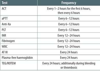
Summary of available coagulation tests is shown in Table 2. The suggested test frequency [1, 31] is shown in Table 3.
TABLE 2
Summary of available coagulation tests
TABLE 3
Frequency of coagulation testing
Tests (aPTT, ACT or anti-Xa) are also performed 4 hours after FFP or antithrombin III (AT III) administration. Recommended targets [31]:
anti-Xa activity 0.35–0.7 IU mL–1;
aPTT 1.5–2.5 times baseline;
ACT, most often recommended at 180–220 seconds, which may vary by device.
Where significant discrepancies between results are found, potential causes should be analysed in detail.
In selected situations such as heparin-induced thrombocytopenia (HIT), heparin resistance, or circuit thrombosis despite adequate anticoagulation, bivalirudin may be an alternative [36]. Current evidence is insufficient to recommend bivalirudin as first-line therapy.
Bivalirudin is a direct thrombin inhibitor that binds both free and clot-bound thrombin. It is meta- bolised mainly by proteolytic enzymes, with 20% renal excretion. Paediatric half-life is 15–42 minutes but is prolonged in renal failure. Advantages include a predictable effect and stable anticoagulation. Disadvantages include limited experience, high cost, no specific antidote, and no specific monitoring assay. Treatment is generally monitored by aPTT, target 60–90 seconds.
Strategy for blood and blood-product transfusion in children on VV ECMO
According to ELSO guidelines [34] and the Pediatric Extracorporeal Membrane Oxygenation Anticoagulation CollaborativE (PEACE) Consensus Conference [35], transfusion of blood and blood products in ECMO patients should be restricted.
Red blood cell concentrate: No clear recommendation has been issued on the optimal haemoglobin level in a child on ECMO. The 2021 ELSO guidelines [31] suggested Hb > 7–9 g dL–1, except in neonates, in children with cyanotic congenital heart disease and in patients with persistent hypoxaemia despite increased ECMO blood flow. This recommendation was not upheld by the PEACE Consensus Conference; in the absence of bleeding, the transfusion decision depends on the clinical picture and on overall oxygen consumption and delivery rather than haemoglobin alone.
Plasma: There is no benefit from prophylactic transfusion of plasma or clotting-factor concentrate when international normalized ratio (INR) < 1.5.
Platelet concentrate: There is no benefit from prophylactic platelet transfusion when platelet count is > 100 000 µL–1.
Fibrinogen: In patients with fibrinogen < 1 g L–1, prophylactic cryoprecipitate and/or fibrinogen concentrate may be considered to prevent bleeding.
Antithrombin III: There are no data on routine AT III administration in ECMO patients. When a therapeutic effect cannot be achieved despite escalating heparin doses, AT III may be considered. Target activity: AT III > 50–80%. After administration, coagulation tests and clinical assessment for bleeding or thrombosis should be repeated.
Bleeding prophylaxis: Topical haemostatic agents are recommended for bleeding prophylaxis.
Management for procedures with low bleeding risk on VV ECMO
These are procedures in which bleeding is easy to recognise and control:
Management for procedures with high bleeding risk on VV ECMO
Consider antifibrinolytics before the procedure and for at least 24 hours afterwards.
Maintain platelets > 100 000 µL–1 and fibrinogen > 150 mg dL–1.
Use topical haemostatic agents.
Consider reducing or pausing anticoagulation, depending on the procedure type and site, the possibility of surgical haemostasis, the patient’s condition and the state of the circuit.
Recombinant factor VII (rFVIIa) is not recommended because of the high thrombotic risk, except for life-threatening bleeding refractory to transfusion.
Management of haemorrhage on VV ECMO
Maintain:
In active haemorrhage, consider reducing or pausing anticoagulation.
In bleeding refractory to transfusion, antifibrinolytics and reduced/paused anticoagulation, consider prothrombin complex concentrate.
In massive haemorrhage, activate a massive trans-fusion protocol with balanced resuscitation using blood and blood products.
In massive haemorrhage with signs of consumptive coagulopathy, evaluate all potential causes including the state of the circuit, patient thrombosis or the development of disseminated intravascular coagulation.
Nutritional support
Critically ill children on ECMO develop metabolic disturbances driven by a generalised inflammatory response and hypermetabolism, with markedly increased energy requirements, deficits and losses of protein and micronutrients, renal dysfunction and electrolyte disturbances. ECMO patients have an increased protein requirement, as protein catabolism is more pronounced than in any other critically ill group. Many are already malnourished before ECMO begins. In paediatric ECMO patients, malnutrition is an independent risk factor for inhospital mortality, morbidity and prolonged ECMO duration [37].
Nutritional status should be assessed at the start of ECMO and monitored throughout: body weight, height-for-weight, anthropometric measurements and albumin levels.
Basal energy requirement should be calculated using the Schofield equation (Table 4) [38]. Total requirement depends on the underlying disease and treatment (sedation, mechanical ventilation) and may exceed the basal requirement severalfold (for example, by 300% in sepsis) [39].
Minimum protein intake should be 1.5 g kg–1 d–1.
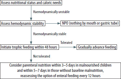
Enteral nutrition should be the primary route; in addition to delivering energy and nutrients, it supports gut function (gut-associated lymphoid tissue [GALT] and the intestinal microbiome) and reduces the risk of bacterial translocation. Critical illness is, however, often accompanied by feed intolerance: gastroparesis, gastro-oesophageal reflux, impaired bowel motility, intestinal ischaemia, paralytic ileus and dysbiosis, leading to vomiting, gastric residuals, diarrhoea or constipation that prevents initiation or continuation of enteral nutrition. Strategies to support gastrointestinal function include prokinetics, minimising sedation, elevating the upper body by 30–45°, post-pyloric feeding tubes, formula changes, laxatives, and switching from continuous infusion to small-bolus delivery.
Implementation of nutritional support is summarised in Figure 1.
Sedation
The aim of sedation is to ensure patient comfort and safe operation of the VV ECMO circuit. General principles do not differ from other paediatric ICU settings. Sedation depth should be monitored with the COMFORT-B scale at least twice daily [39]. Analgosedation is provided as an opioid infusion (morphine, fentanyl, sufentanil), with benzodiaze-pines and/or dexmedetomidine or clonidine. Non-opioid analgesics (paracetamol and metamizole) should be added. Ketamine infusion is also recommended when deeper sedation is required. Because of the risk of propofol infusion syndrome, propofol infusions of more than 48 hours or at doses above 4 mg kg–1 h–1 are not recommended [39].
Maintaining the patient’s own respiratory drive appears beneficial, as it supports pulmonary rehabilitation. Excessive respiratory effort, however, may increase the transpulmonary pressure gradient and lead to patient self-inflicted lung injury (P-SILI) [40].
In specific situations such as hypermetabolic states with high oxygen consumption, neuromuscular blocking agents may be useful, with monitoring of the degree of blockade. Instrument-based methods of sedation depth (e.g. BIS, SedLine) are available but have no firm paediatric recommendations.
Analgosedation is often associated with impaired gastrointestinal function, disuse muscle atrophy, tachyphylaxis and dose escalation, all of which directly increase the risk of delirium and withdrawal during weaning. These considerations have led to the concept of extubating patients and managing them on ECMO without mechanical ventilation, thus avoiding deep sedation (“awake ECMO”). This is particularly relevant for patients expected to require prolonged therapy, such as those awaiting lung transplantation. ELSO Registry data for 2018–2022 show that 4.8% of paediatric ECMO patients were managed while breathing spontaneously. The non-intubated group had shorter ICU stays and faster mobilisation and rehabilitation. However, mortality was higher in this group, and the authors note that an association between extubation and mortality cannot be excluded. At this stage, extubation of patients on ECMO is recommended only in centres with extensive experience [41].
Fluid management
Fluid overload of 10% during ECMO may be associated with longer therapy and higher mortality, and prevention or correction of overload is an important factor in improving pulmonary function and reducing ECMO duration [42–44]. Fluid overload is calculated as the ratio of daily cumulative fluid balance to admission body weight:
% fluid overload = (fluid balance / admission body weight) × 100
Principles of fluid management in ECMO patients [45]:
Fluid balance should be reviewed at least every 12 hours.
Aim for a negative fluid balance.
Restrict fluids to those essential, and concentrate solutions.
Maintain urine output sufficient to achieve the target balance.
Consider albumin to maintain serum albumin > 2.5 g dL–1.
If overload develops despite fluid restriction and diuretics, start renal replacement therapy (RRT) [46].
RRT may be delivered as continuous renal replacement therapy, or less commonly as peritoneal dialysis or intermittent haemodialysis. Continuous techniques (haemofiltration or continuous venovenous haemodiafiltration [CVVHDF]) are used most often because they allow stable, controlled fluid removal with haemodynamic stability. RRT can be connected to the ECMO circuit in three ways: a separate venous access, in-line addition of a haemofilter to the ECMO circuit, or integration of a CVVHDF device into the circuit.
According to ELSO recommendations, the CVVHDF device is connected to the venous (return) line [47]:
blood is drawn after the pump, either before or after the oxygenator;
blood is returned before the oxygenator.
Early initiation of RRT may favourably affect duration of ECMO support and survival [45, 47].
Ventilation
Extracorporeal oxygenation minimises the risks of barotrauma, volutrauma, atelectotrauma and biotrauma associated with mechanical ventilation.
Suggested ventilator settings: FiO2 < 50%, PIP < 25 cmH2O, PEEP 5–15 cmH2O, respiratory rate 10–20 min–1, inspiratory time 0.8–1 second.
Ventilation should take account of the patho-physiology of the underlying disease; for example, inspiratory time should be shorter in status asthmaticus.
Pneumothorax and any pleural fluid or blood should be drained.
Airway clearance should be performed meticulously, including bronchoscopic suction where indicated.
Recruitment manoeuvres are not recommended.
Other aspects of care for the ECMO patient [1]
Rehabilitation: physiotherapy should be started as early as possible after haemodynamic stabilisation and once ventilator parameters (especially PEEP) are stable, following the principle of early mobilisation. Identify patients suitable for passive exercises and those who can engage in active rehabilitation.
Eye care. Patients unable to close their eyes are at risk of keratopathy with ulceration and infection. The eyes should be examined at least every 12 hours. Lubricating drops and protective covers to prevent drying are recommended.
Pressure-ulcer prevention.
Oral hygiene.
Infection control: there is no rationale for routine prophylactic antibiotic therapy.
COMPLICATIONS OF ECMO
VV ECMO is a highly specialised therapy used predominantly in patients with severe respiratory failure. Survival in this group exceeds that of VA ECMO, but the therapy still carries substantial risks of specific mechanical and haematological complications. Complications can be grouped as haematological, neurological, infectious and equipment-related (Table 5). Survival at 24 hours after decannulation and to hospital discharge is 74.5% and 63.8%, respectively [49]. The median duration of therapy is 188 hours [49].
TABLE 5
Incidence of selected complications per 1,000 hours of paediatric VV ECMO
Bleeding
Incidence
Bleeding is a common complication of paediatric VV ECMO; reviews indicate significant bleeding in up to about 40–50% of children. The most frequent sites are the surgical wound, cannulation site, CNS, lungs and gastrointestinal tract [50, 51].
Monitoring
Routine measurements: complete blood count (platelets), PT/INR, aPTT, ACT, fibrinogen, D-dimers, anti-Xa [35].
During bleeding, fibrinogen should be monitored and maintained at ≥ 150 mg dL–1 (≥ 1.5 g L–1) [52].
Inspect cannulation sites, drains, surgical sites and assess for internal bleeding (lungs, gastrointestinal tract).
Management
In active bleeding: correct haemostasis (replace fibrinogen, platelets and clotting factors) and modify the anticoagulation strategy (lower dose, change of agent).
In difficult-to-control bleeding: consider replacing components of the VV ECMO circuit, or reducing flow if feasible. Detailed management is discussed by Rintoul et al. [53].
The transfusion strategy (red cells, platelets, cryo-precipitate) should take into account the risk of thrombosis.
Iatrogenic blood loss from laboratory sampling should be minimised.
Thrombosis/clotting
Incidence
Thrombosis within the VV ECMO circuit or in the patient is reported in approximately 20–30% of cases [35, 50].
Management
If a thrombus is detected in the circuit: replace components (oxygenator, pump or lines) promptly; review flow settings and cannula position.
Adjust anticoagulation: if clots occur on a “standard” dose, consider increasing anticoagulation, changing the agent or modifying the haemostasis protocol [54].
Haemolysis
Incidence
Haemolysis is among the more common mechanical complications of paediatric VV ECMO and may lead to renal injury and increased risk of coagulation-related complications. Clinically significant haemolysis is defined as plasma-free haemoglobin (pfHb) above 0.5 g L–1 or lactate dehydrogenase more than three times the upper limit of normal. Up to 43.5% of patients develop severe haemolysis (pfHb ≥ 1.0 g L–1). Dalton et al. [51] found that higher haemoglobin and more frequent red cell transfusions were associated with increased haemolysis, whereas well-managed anticoagulation was protective. Monitoring of haemoglobin, pfHb and signs of haemoglobinuria is central; in rising haemolysis, mechanical causes such as thrombus in the pump or oxygenator, abnormal flow or suboptimal cannula position must be excluded. Correction of pump and flow settings and, when necessary, replacement of circuit components (oxygenator/ pump) are the mainstays of management [55].
Neurological complications
Incidence
Seizures and other neurological complications occur in around 15% of cases; the majority are sub-clinical. They are more frequent after extracorporeal cardiopulmonary resuscitation (ECPR) and in children supported on VA ECMO [56].
Management
On detection of intracranial haemorrhage or stroke: collaboration with neurology/neurosurgery; consideration of changing or reducing anticoagulation; clinical decisions on continuation of VV ECMO in light of neurological prognosis.
Prevention: avoid aggressive mechanical ventilation, control intracranial pressure, maintain adequate anticoagulation.
Infections
Incidence
VV ECMO-related infections (cannulation-site infection, bacteraemia) occur in approximately 10–20% of patients. Risk rises with duration of therapy. Infection is associated with an approximately twofold increase in mortality. Candida spp. are notable pathogens [9].
Management
Routine antibiotic prophylaxis during VV ECMO remains controversial; current ELSO guidance does not recommend it routinely [1]. If infection is suspected, promptly initiate empirical antibiotic therapy, obtain microbiological samples, and consider cannula relocation or replacement if the insertion site is suspected to be the source of infection.
Strict aseptic technique during all VV ECMO handling, minimisation of unnecessary circuit changes, and prompt termination of therapy when possible.
Cannulae and recirculation
Incidence and significance
A specific technical issue in VV ECMO is recirculation: direct return of oxygenated blood into the drainage limb. Recirculation depends on the relative position of drainage and return cannulae, the flow rate and the patient’s blood volume. High recirculation (> 25–30%) reduces effective oxygen delivery and manifests clinically as low systemic saturation despite normal VV ECMO parameters. Small vessel diameter is a further technical limitation; in children weighing less than 15 kg, femoral cannulation is discouraged. The risk of cannula kinking with small sizes should be considered, as should the lesser stability of such cannulae, given the short insertion path and poorly developed subcutaneous tissue [1].
Management
Secure cannula fixation to prevent displacement. If recirculation is suspected, reposition the cannula, change configuration (e.g. to a dual-lumen cannula or another arrangement) and ensure adequate flow [57].
Equipment failure
Incidence
Failure of the oxygenator, pump or lines can lead to haemolysis and impaired gas exchange and, consequently, worse outcomes. Paediatric data indicate that circuit component repla cement is required in approximately 10–15% of thera- pies [58].
Management
When deterioration is detected, a prompt decision should be made to replace the affected component (oxygenator/pump/circuit).
Regular technical checks in cooperation with the perfusionist, scheduled circuit-change protocols based on time in service, and planned, controlled component replacement to avoid sudden failure at a critical moment.
WEANING AND DECANNULATION FROM VV ECMO
Timely termination of extracorporeal mechanical support is as important as its initiation. VV ECMO should be discontinued when progressive recovery and return of native lung function can be demonstrated, the child tolerates gradual reduction of VV ECMO support, and reduction does not cause clinically meaningful deterioration in gas exchange [1].
ECMO is an aggressive therapy with potential for serious complications. Duration should be as short as possible, and every ECMO patient should be assessed daily for response to therapy and readiness for weaning and continuation of care with less invasive techniques (e.g. mechanical ventilation) [59].
When optimal recovery of pulmonary, cardiac and other organ function has been achieved, a “weaning trial” can be performed to obtain as much information as possible on the likelihood of successful VV ECMO discontinuation. The trial is a diagnostic, not therapeutic, intervention. It allows determination of the patient’s readiness to continue care without VV ECMO.
Definition
Successful termination of VV ECMO is defined as 48-hour survival of the child after extracorporeal mechanical gas-exchange support is stopped, without need to restart extracorporeal therapy [1].
Criteria for termination of VV ECMO therapy
Weaning from veno-venous support is one of the most difficult aspects of ECMO care. There are three categories of decision to terminate extra-corporeal support:
Clinical improvement, with reasonable confidence that ECMO can be successfully terminated.
Low likelihood of successful termination, but ECMO cannot be continued because of complications or high risk thereof.
Termination because of a fatal, incurable disease or severe complications (compassionate withdrawal).
In the first case, before terminating ECMO, a decision should be made whether the patient will be re-eligible for ECMO in the event of failure to wean, and under what circumstances.
In the third case, ECMO is withdrawn as part of palliative care after discussion with the family, in the last phase of life, when withdrawal allows natural death from the underlying disease.
In the second case, short- and long-term outcomes are harder to predict. Switching to less invasive treatment may worsen organ function despite intensive mechanical ventilation and pharmacological circulatory support, while continued ECMO carries an excessive risk of mortality or major morbidity. Before disconnection, a decision should be made on whether, and under what circumstances, the patient will be eligible for re-cannulation onto ECMO.
Table 6 lists the factors to consider before deciding to terminate ECMO [1].
TABLE 6
Factors considered in the decision to terminate VV ECMO
There are no fixed time limits or a single universal weaning algorithm. Each patient has an individual algorithm dictated by the clinical context; this should be recognised and the weaning trial tailored accordingly to maximise the chance of successful liberation.
Weaning trial
ELSO Registry data and case series indicate growing use of VV ECMO in children with respiratory disease [48]. There is also some case-series and registry evidence that survival may be better with VV than VA ECMO. VA ECMO is sometimes chosen at the outset in the most severe PARDS until improvement allows a switch to veno-venous support.
De-escalation differs between VA and VV ECMO. Weaning from VV ECMO consists of a sweep-gas off trial, the ultimate aim of which is to assess pulmonary function while the ECMO circuit is not contributing to gas exchange. The sweep gas is therefore stopped while blood flow through the ECMO circuit is maintained. During the trial, the patient is on full lung-protective mechanical ventilation, with monitoring of gas exchange, respiratory effort and haemodynamics [60].
The sweep-off trial begins with stepwise reduction of sweep-gas FiO2 to 0.21 over 20–30 minutes. The 21% oxygen sweep mixture is then maintained for a further 20 minutes (until blood colour in the circuit equalises) while the child is observed for changes in SpO2, respiratory rate, heart rate, blood pressure or rising agitation. After another 20 minutes, the sweep-gas flow is turned off. Gas exchange will then occur only through the lungs. Observation usually lasts 2–6 hours. Adrenaline and inhaled nitric oxide should be on hand in case of pulmonary vasoconstriction and a rise in pulmonary arterial pressure. As sweep-gas flow and FiO2 are reduced, an arterial blood gas should be obtained every hour, sampled within 30 minutes of each change, to assess oxygenation and ventilation [61]. Sudden cessation of sweep flow lowers a relatively high venous saturation and raises PaCO2; this can substantially disturb V/Q matching, abruptly lower saturation, raise pulmonary vascular resistance and cause the weaning trial to fail. Sequential reduction of sweep-gas FiO2 before complete cessation is therefore both rational and physiologically grounded.
The child’s own lungs should be able to support 50–80% of total gas exchange. In severe lung disease, weaning from ECLS may begin when the native lungs reach 80% of oxygen delivery and respiratory mechanics show substantial improvement [62]. One method of assessing lung recovery is the oxygen challenge test (OCT). ECMO parameters are held constant while FiO2 is increased to 1.0; the resulting increase in PaO2 is then assessed. Higher absolute PaO2 or a larger ΔPaO2 suggests adequate recovery and the possibility of weaning. Evidence is currently insufficient to define threshold PaO2 or ΔPaO2 values for initiating weaning [62].
Available evidence does not support superiority of any specific ventilator mode during VV ECMO weaning [62]. Ventilator settings should, however, be modified to accept PaO2 values of 60–80 mmHg and PaCO2 of 40–60 mmHg, allowing lower airway pressures and lower FiO2 [1].
If ventilatory or haemodynamic problems develop at any point, the weaning trial should be aborted immediately and full ECMO support restored. SpO2 < 88% indicates failed weaning.
If the patient is judged not yet ready for liberation, or the team is not available to decannulate, the circuit FiO2 should be returned to 100% and sweep gas to the previous setting; mechanical ventilation may be continued or reduced pending decannulation.
Decannulation
Decannulation should ideally be performed in the morning to allow full management of any deterioration. Cannula removal is usually performed by the cardiac surgical team in the paediatric intensive care unit. A plan for managing deterioration after disconnection should be agreed in advance.
Anticoagulation is maintained up to the point of decannulation. In patients with a percutaneous catheter, heparin is stopped one hour before catheter removal. Thirty minutes before cannula removal, a prophylactic dose of an antibiotic such as cefazolin (30 mg kg–1) is given.
Complications after ECMO termination
Complications may occur after successful disconnection even in patients who tolerate the first few hours well. The most commonly reported include bleeding from cannulation sites, limb ischaemia, ischaemia of organs such as the kidneys, liver and nervous system, neurological complications and infections [62].
Risk factors for death after successful ECMO termination include prolonged ECMO support with complications, irreversible organ injury, low pH with high lactate levels, and advanced organ failure [62].
SUMMARY
Extracorporeal cardiopulmonary support should be considered in children with severe respiratory failure when conventional methods fail to provide adequate gas exchange, when they require potentially harmful ventilator settings, or when severe barotrauma occurs. VV ECMO is currently recommended as a treatment option for PARDS, particularly after optimisation of conventional management, including LPS, prone positioning and neuromuscular blockade. VV ECMO may also be used as perioperative support to enable selected procedures such as airway surgery. Because of the small number of methodologically robust studies, current ELSO and PALICC-2 recommendations are general in nature and emphasise the need for further, well-designed studies in this area. The present document synthesises current recommendations with the aim of improving outcomes in paediatric patients with PARDS in Poland.
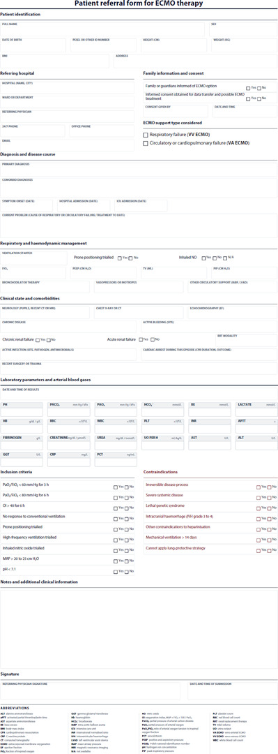
The patient referral form for ECMO is provided in Appendix 1.