
Percutaneous arterial cannulation is one of the most frequently performed invasive procedures in critically ill patients. The indications for arterial cannulation are continuous arterial pressure monitoring, repeated arterial blood sample collection, and hemodynamic monitoring using transpulmo-nary thermodilution [1]. In such cases, the radial artery is usually cannulated, however the common femoral, brachial, and axillary arteries can be used when the radial artery is inaccessible [2].
Femoral access for arterial line placement is frequently used as an alternative site when the radial artery cannot be cannulated, for example in the case of excessive arterial vasoconstriction or circulatory shock. On the other hand, few prospective clinical trials have been carried out on femoral arterial access performed in the intensive care unit (ICU). The vast majority of studies regarding femoral artery catheterization have been conducted in the fields of interventional cardiology and endovascular surgery. In contrast, many trials have focused on radial artery catheterization in critically ill patients [3, 4].
The femoral artery in the ICU is a common alternative to the radial approach for arterial line placement and may even be the first choice for arterial catheterization in transpulmonary thermodilution. Of note, the indications for an immediate arterial catheter placement in critically ill patients may be revised in the future, especially in view of the paper by Muller et al. [5]. However, a large percentage of patients in shock will still require large artery catheterization, notably because of advanced hemodynamic monitoring. Another reason for large artery cannulation is the common occurrence of the central-to-peripheral arterial blood pressure gradient [6]. When this occurs, radial artery pressure monitoring may underestimate arterial pressure, resulting in administration of increased doses of vasoactive agents [7]. It seems that modern intensive care requires a personalized approach to hemodynamic monitoring. This means that certain patients may be monitored non-invasively while others need large artery (e.g. femoral artery) cannulation and advanced hemodynamic monitoring.
This narrative review therefore aims to synthesize the current evidence regarding the efficacy and safety of common femoral artery (CFA) cannulation for arterial pressure monitoring in critically ill adult patients and discusses the most important practical issues related to this access. Technical aspects of CFA cannulation in intensive care are also presented. To the best of our knowledge, there has been no review paper published on cannulation of the femoral artery for arterial line placement in ICU patients.
METHODOLOGY
A literature search was performed to identify studies conducted among critically ill patients who had had a femoral artery cannula inserted percutaneously for arterial pressure measurement. The studies identified had to report the number of cannulated patients, describe the procedure, state the number of days of catheter maintenance, and describe any peri-procedural complications. Three databases (Medline, Scopus, and Web of Science) were screened from inception to 10 December 2025. The search was restricted to studies conducted in ICUs and written in English. The following search strategy was used: “critically ill” or “intensive care” or “critical care” or “arterial pressure” and “femoral artery or common femoral artery” and “catheter* or cannul*”. Abstract screening, followed by a full-text assessment, was performed by two blinded researchers (RG and TK). Any discrepancies were resolved by discussion.
SEARCH RESULTS
The search yielded 3196 studies. After excluding 1186 duplicates, 2010 records were screened, and nine studies were included that had been conducted on critically ill patients who had catheters inserted for invasive arterial pressure measurement. Table 1 presents a summary of the data of the studies identified. The studies were not consistent regarding the methodology, cannulation technique, reported outcomes, or complications, and more than 50% were conducted over 40 years ago.
TABLE 1
Studies on percutaneous cannulation of the femoral artery for measurement of arterial pressure in critically ill patients
In the majority of papers, the femoral artery was cannulated using only anatomical landmarks with palpation guidance [8–14]. Among these studies, only Thomas et al. [13] reported an overall success rate, which was 99%, while Belda et al. [11] reported a first-pass success rate of 87.4%. Two randomized controlled trials (RCTs) examined ultrasound-guided techniques. Gutte et al. [15] compared ultrasound (US)-guided CFA cannulation with a palpation-based technique and found that ultra-sonography yielded a higher first-pass success rate and shorter cannulation time, although complications were not reported. In a non-inferiority RCT, Gawda et al. [16] compared US-guided CFA cannulation with US-guided infraclavicular cannulation of the axillary artery. The first-pass success rate for CFA cannulation was 74.1%, and the most common peri-procedural complication was periarterial blood extravasation, occurring in 20.4% of patients.
EVIDENCE FROM THE NON-ICU SETTING
The vast majority of contemporary prospectively conducted trials regarding US-guided CFA cannulation originate from cardiac catheterization laboratories where various (including large bore) catheters/ sheaths were inserted for coronary angiography, transcatheter valve replacement, or implantation of mechanical circulatory support devices. Considering that the procedures in these trials differ from arterial line placement for invasive arterial pressure measurement in the ICU, the conclusions based on these studies should be drawn very cautiously for the population of critically ill patients. Additionally, only studies in which coronary angiography/ coronary interventions were performed using thin catheters/sheaths (equal to or lower than 5 Fr in diameter) should be considered.
Most of the RCTs concerning CFA cannulation for coronary intervention support the use of ultra-sound guidance, which is associated with a better first pass success rate and lower numbers of arterial puncture attempts and peri-procedural complications. However, Dudeck et al. [17] proved that, compared to palpation-based methods, US-guided cannulation is only better in obese patients and in patients with a weak arterial pulse. Dudeck et al. [17] used sheaths for angiography that were no more than 5 Fr in size. Seto et al. [18] compared US-guided CFA retrograde cannulation for sheath insertion (mean size 5.6 Fr) with fluoros-copy guidance and pulse palpation. The US-guided method demonstrated a higher first-pass success rate, lower number of arterial puncture attempts, and fewer complications [18]. In the study by Gedi koglu et al. [19], patients were randomly allocated to a palpation-guided group and US-guided group for sheaths insertion during angiography. No significant differences in cannulation success rate and complications were shown; however, the US-guided method demonstrated a higher first-pass success rate and fewer attempts. No differences were found between US-guided and pulse-based catheterization in a study by Marquis-Gravel et al. [20] in which patients were cannulated for coronary angiography or coronary interventions except for several venipunctures. Jolly et al. [21] compared ultrasound guidance and fluoroscopy for femoral arterial access in coronarography/cardiac interventions with the palpation and fluoroscopy methods. In this bi-center RCT including 621 patients, ultra-sonography reduced the risk of venipuncture and number of attempts, but it did not reduce vascular complications or the risk of bleeding. However, the patients were cannulated with a 6 Fr or larger introducer, and in half of the procedures, a closure device and unfractionated heparin were used.
WHAT ARE THE MAIN FEATURES OF THE FEMORAL ARTERIAL LINE IN INTENSIVE CARE?
Arterial measurement via the femoral artery has several differences compared to the routine measurement using the radial artery.
The values of arterial pressure measured via CFA exceed those in smaller arteries. The so-called “radial-to-femoral” difference has been examined in several trials [22–25]. Additionally, a recent meta-analysis by Hasegawa et al. [26] confirmed that the values of systolic blood pressure in the femoral artery are slightly greater than those in the radial artery. Arterial pressure measured in the CFA may influence treatment decisions in critically ill patients. This is because the threshold values of arterial pressure in some conditions (e.g. in septic shock) can be achieved with lower doses of catecholamines. The radial-to-femoral difference also has clinical implications regarding arterial pressure measurement during liver transplantation or cardiothoracic surgery [27, 28].
Second, the catheters inserted into the CFA are more durable compared to those inserted into the radial artery [29, 30]. This feature may be practically useful when the patient’s stay in the ICU is predicted to be longer. In addition, patients with the catheter placed in the femoral artery can undergo early mobilization [31, 32]. However, a longer period of maintenance is also associated with an increased risk of arterial catheter related bloodstream infection (ACRBSI) [33].
Third, the inguinal area for arterial catheter insertion is regarded as the site of the last choice compared to other areas such as the wrist (for radial artery cannulation) or foot dorsum (for dorsalis pedis artery cannulation). Many studies have confirmed that incidents of ACRBSI are more likely when the catheter is inserted in the femoral rather than the radial or dorsalis pedis arteries [34–36]. Even though this claim was not confirmed in an analysis based on a very large dataset, the inguinal area is still regarded as the location with highest risk for ACRBSI [37].
WHEN SHOULD THE FEMORAL APPROACH BE PREFERRED OVER THE RADIAL ARTERY CANNULATION?
There are possible scenarios in which the femoral artery should be considered as the first-choice option for arterial line placement compared to the radial artery:
CLINICAL ANATOMY
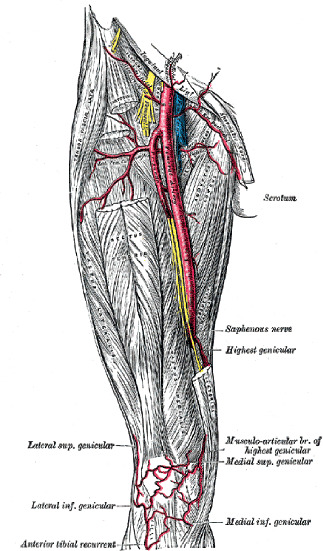
The CFA is a continuation of the external iliac artery and runs from the midpoint of the inguinal ligament (or from the branching of the inferior epigastric artery) to the bifurcation of the CFA and divides into the superficial and deep femoral arteries (Figure 1). Of note, the inguinal crease does not lie directly over the inguinal ligament, which is usually located higher up. The distance between the inguinal crease and inguinal ligament varies, with an average value of 6.7 cm, and the bifurcation of the CFA in the majority of patients (76.5%) lies over the inguinal crease [38].
FIGURE 1
Human anatomy of the femoral vessels. The diagram is reproduced from Gray’s Anatomy, 1918, 20th US edition
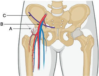
The mean diameter of the CFA in healthy males and females is 9.8 mm and 8.2 mm, respectively [39]. The depth of the CFA usually does not exceed 2 cm, but it can vary depending on the patient’s physical status, especially the thickness of the subcutaneous tissue [40, 41]. In a patient who is lying on their back, most of the CFA is located above the head of the femoral bone. The bifurcation of the CFA is usually located below the caudal border of the head of the femoral bone (Figure 2).
FIGURE 2
Mutual position of the common femoral artery, the inguinal crease, the inguinal ligament, and the head of the femoral bone. A) Inguinal crease. B) Common femoral artery. C) Inguinal ligament. Created using BioRender.com
The femoral vein lies medially and posteriorly to the CFA, and the femoral nerve lies laterally to the CFA (Figure 1). The mutual location of the CFA and femoral vein changes slightly depending on the distance from the inguinal ligament. The vessels are more distant at the beginning of the CFA; however, more caudally, the femoral vein lies closer to and posterior to the CFA [42]. In older patients, the CFA is often affected by significant atherosclerosis, thus precluding efficient catheterization [43].
THE MAIN PRINCIPLES OF CANNULATION
The Seldinger technique (catheter-over-guide-wire) is the most commonly used method of femoral artery cannulation in intensive care [44]. For arterial pressure measurement, a retrograde cannulation is performed in which the tip of the catheter is inserted in the opposite direction to the arterial blood flow. Single-lumen catheters are typically thin (18 or 20 gauge in diameter), 10 to 20 cm in length. The entire procedure should be performed under sterile conditions using a sterile set with a catheter and a sterile dressing for the patient. The vessel should be punctured below the inguinal ligament and over the bifurcation of the CFA.
LANDMARK-BASED ACCESS
Traditionally, anatomical points with pulse detection over the CFA were used for catheterization in the pre-ultrasound era. When this technique is mentioned throughout the paper, the term “landmark-based” is used in this regard. In the landmark-based approach, the operator identifies the course of the inguinal ligament and then locates the CFA below the mid-point of the inguinal ligament using the fingertips of the non-dominant hand in order to detect the most intensive pulsation over the artery [45]. Simultaneously, the needle with the attached syringe is held in the dominant hand. The skin over the CFA is punctured by the needle approximately 2–3 cm below the inguinal ligament. The needle is then introduced at an angle of around 45 degrees in the cranial direction towards the artery along the long axis of the artery. During needle insertion, negative pressure in the connected syringe should be generated by withdrawing the plunger while inserting the needle. When the needle punctures the arterial wall, arterial blood is aspirated. The syringe is then disconnected, the needle is held in the non-dominant hand, and after confirmation of free pulsatile blood outflow, the guidewire is inserted through the needle into the lumen of the CFA. In the next step the needle is removed from the artery, while the guidewire is still in the artery. Immediately afterwards, the cannula is introduced over the guidewire into the lumen of the CFA, and when the cannula has been completely inserted, the guidewire is withdrawn from the artery. In the final step, the catheter is sutured to the skin and a sterile dressing is applied.
ULTRASOUND-GUIDED ACCESS
In accordance with current recommendations, cannulation of the arteries should be performed using real-time ultrasound guidance [46, 47]. US-guided cannulation of the CFA can be performed using the out-of-plane or in-plane technique.
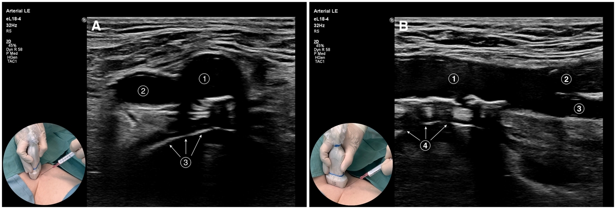
Out-of-plane cannulation occurs when the needle is inserted perpendicular to the plane of the ultra-sound beam. The CFA in this method is visualized in the short-axis view (see Figure 3A).
In-plane cannulation occurs when the needle is inserted in-plane of the ultrasound beam. Practically, the CFA is visualized in the long-axis view (see Figure 3A).
FIGURE 3
Different methods of US-guided cannulation of the common femoral artery (CFA). A) Short-axis view of the femoral vessels and the out-of-plane approach to the CFA (circular photo on the left): 1 – CFA, 2 – femoral vein, 3 – head of the femoral bone. B) Long-axis view of the CFA with its bifurcation and the in-plane approach to the CFA (circular photo on the left): 1 – CFA, 2 – superficial femoral artery, 3 – deep femoral artery, 4 – head of the femoral bone
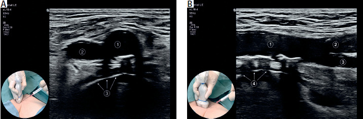
The out-of-plane approach with the short-axis view of the artery is more usually recommended, because the artery is easier to visualize, especially in obese patients [46]. The main advantage of this approach is that it is able to visualize the surrounding structures such as the femoral vein and femoral nerve. The out-of-plane approach also enables the operator to use a steeper angle (around 45o) during the needle insertion.
The in-plane technique with a long-axis view is more difficult to perform than the out-of-plane method with a short-axis view, because of the need for precise hand coordination in order to keep the needle within the ultrasound beam. The main benefit of this method is being able to visualize the entire needle with its tip during insertion.
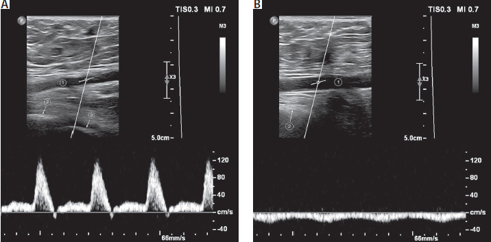
Distinguishing the femoral artery from the fe-moral vein can be challenging using the in-plane technique. This is because both vessels may appear similar on the screen and can rarely be visualized simultaneously in the long axis-view. Pulsed-wave Doppler is a recommended tool for femoral vessel identification in such cases as shown in Figure 4.
FIGURE 4
Pulsed-wave Doppler is used to distinguish the common femoral artery (CFA) from the femoral vein. A) 1 – CFA, 2 – hip socket, 3 – head of the femoral bone. B) 1 – Femoral vein, 2 – head of the femoral bone
The US-guided procedure can be divided into a few consecutive steps:
Before starting the procedure, pre-scanning of both inguinal areas is crucial to assess the suitability of conditions for US-guided cannulation. The operator should choose the CFA, which is wider, shallower, and without the presence of significant atherosclerosis.
Catheterization is performed under sterile conditions. The puncture site should be prepared with a sterile alcohol-based chlorhexidine solution. The operator should wear a sterile gown and sterile gloves, and the patient should be fully draped with sterile sheets, with the ultrasound probe and cable being completely covered with the sterile sleeve.
The CFA should be examined with ultrasound several times (using short-axis and long-axis view) from the inguinal ligament caudally to the bifurcation of the CFA. The head of the femoral bone should also be visualized to confirm that the CFA is located just above the head of the femoral bone, thus ensuring an appropriate puncture site (see Figure 3).
The transducer should be held with the non-dominant hand, and the needle with the attached syringe held in the dominant hand. The needle should puncture the skin just in front of the probe. Once the needle can be visualized on the screen, it is inserted towards the wall of the artery at an angle of approximately 45 degrees. The artery should be punctured in the mid-portion of the CFA, over the head of the femoral bone and below the inguinal ligament.
When the tip of the needle is visible inside the artery, the syringe is disconnected and the guide-wire is advanced through the needle into the artery. The intra-arterial position of the guide-wire is then confirmed using ultrasound. After ensuring that the guidewire is in the right position, the needle is withdrawn, the guidewire is left inside the CFA, and the cannula is inserted over the guidewire into the lumen of the artery. After confirming the intra-arterial position of the cannula, the guidewire is withdrawn from the CFA.
Finally, the catheter is secured to the skin with sutures. Although some recommendations advise using a sutureless dressing for central venous catheters, their use of such dressings has never been assessed for arterial catheters [48].
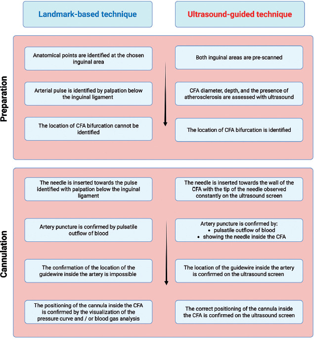
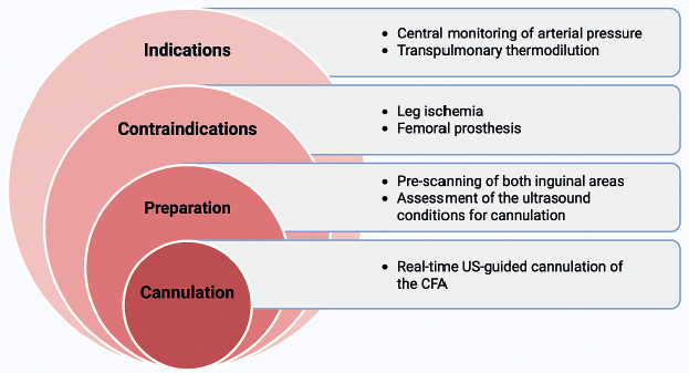
The US-guided approach is different from the landmark-based technique, as shown in Figure 5. The main advantages of the US-guided technique compared to the landmark-guided method are as follows: 1) the best CFA can be selected for cannulation, 2) the presence and the size of the intra-arterial atherosclerosis can be assessed, 3) the CFA can be distinguished from the femoral vein, and 4) the CFA bifurcation level is simple to identify. In daily practice, several factors need to be considered during arterial line placement via the femoral route with the US-guided cannulation, as shown in Figure 6.
PRACTICAL TIPS ON CFA CANNULATION
To minimize the probability of post-procedural hemorrhage, it is important not to introduce the dilator (when applied) into the lumen of the artery using the Seldinger technique. In landmark-based cannulation this can be difficult, but even in US-guided cannulation the dilator cannot be visualized on the screen. To prevent dilator insertion into the artery, the operator should assess the depth of the CFA during the pre-scanning step and prevent the dilator being advanced to a depth greater than the CFA location.
The procedure can be especially challenging in obese patients when the artery lies at a greater depth. In such cases, the operator can shorten the distance from the skin to the artery by applying compression on the inguinal area at the cannulation side with the non-dominant hand. However, special attention should be paid to maintain constant compression when the needle is placed in the artery to prevent inadvertent withdrawal of the needle from the vessel.
The guidewire should be inserted into the CFA only when: 1) there is free pulsatile blood outflow from the artery, and 2) there is no resistance during guidewire insertion. The aim of these prerequisites is to prevent potential CFA dissection by the guidewire inserted into the arterial wall instead of the lumen of the artery. This is especially important during landmark-based cannulation when an incorrectly inserted guidewire cannot be visualized on the screen.
The CFA is only approximately 4 cm long; therefore, good visualization can be challenging during the procedure. As the linear transducer has a breadth of usually around 5 cm, the probe needs to be partially positioned over the inguinal ligament if the long-axis view is used during cannulation.
It is important to minimize the time after withdrawing the needle and before cannula insertion in order to prevent periarterial hematoma caused by blood extravasation. A hematoma forms in such cases because the hole in the artery wall caused by the needle puncture is greater than the diameter of the guidewire. When the arterial pressure is high, bleeding can occur around the guidewire through the puncture hole in the artery.
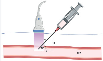
In some circumstances (e.g. morbid obesity) the CFA can be located at such a depth that the length of the needle needs to be carefully checked to ensure that the artery can be reached during the procedure. The Pythagorean theorem is helpful in such a case because the needle is inserted at an angle of 45 degrees, as shown in Figure 7.
FIGURE 7
Example of the use of the Pythagorean assertion in percutaneous cannulation of the common femoral artery (CFA). The depth of the CFA is depicted by the dashed vertical line. The minimum length of the needle (depicted as “c”) inserted at an angle of 45° should be equal to c = a √2 to reach the lumen of the CFA. Created using BioRender.com
CONTRAINDICATIONS
The contraindications for femoral arterial line placement are limited. Absolute contraindications include lack of consent, local infection at the skin puncture site, CFA dissection, and clinically evident symptoms of leg ischemia on the cannulation side.
Relative contraindications include advanced atherosclerosis in the CFA, difficult conditions for ultrasound-guided cannulation (deep location of the artery and/or patient obesity), and severe hemostasis abnormalities.
COMPLICATIONS
Early complications can be divided as follows: 1) typical complications of arterial cannulation, 2) complications linked specifically to arterial cannulation in the inguinal area. Several possible peri-procedural complications associated with arterial cannulation include perivascular hematoma, artery wall dissection, puncture of the opposite wall of the artery, and puncture/cannulation of the adjacent vein [49]. More specific complications for the cannulation of the CFA are femoral nerve injury, bowel/bladder perforation, retroperitoneal hematoma, pseudoaneurysm, and arteriovenous fistula.
Retroperitoneal hematoma
This complication is rare. The most significant factor influencing the occurrence of retroperitoneal hematoma is puncture of the CFA above the inguinal ligament. The literature reports retroperitoneal hematoma in around 0.5% of patients undergoing coronary interventions [50]. When retroperitoneal bleeding is significant, symptoms of hemorrhagic shock may occur. To establish a correct diagnosis, radiological diagnostic tests (USG, CT) should be performed.
Pseudoaneurysm
Pseudoaneurysm is defined as a breach in the arterial wall with subsequent blood leakage to the surrounding adjacent tissues. This complication is rare, with the reported incidence rate ranging from 0.44% in diagnostic procedures to 8% in procedural catheterization [51]. One of the risk factors for femoral pseudoaneurysm formation is a puncture site below CFA bifurcation [52]. Other factors include high BMI, hemostasis abnormalities, large cannula size, inadequate compression after the procedure, and advanced age [51].
Arteriovenous fistula
The reported incidence of arteriovenous fistula (AVF) after transfemoral cardiac catheterization ranges from 0.6 to 1.8% [53, 54]. There are no data describing this complication among patients treated in ICUs. The most important risk factors for AVF in critically ill patients include systemic anticoagulation, multiple punctures, and puncture site at the bifurcation level (and below) of the CFA [55].
Late complications include leg ischemia on the cannulated side and ACRBSIs. Assessing the prevalence of ischemic complications is difficult since the diagnostic criteria in various studies are based only on researchers’ subjective assessments. In a comprehensive review, Scheer et al. [49] reported a rate of 0.18% of severe ischemic complications after femoral catheterization in 3899 patients. In another large analysis of 4447 patients after femoral arterial line placement, ischemic incidents were observed in 0.09% of patients [56]. In some studies identified in the search conducted for this review, ischemic events were more frequent, with a rate of 3.4% in a study by Russell [8]. In contrast, no cases of femoral artery thrombosis and lower limb ischemia were noted in a study on 2318 patients in whom femoral arterial catheter was inserted before induction of anesthesia [14]. However, it should be noted that in the latter study, patients with any lower limb atherosclerotic disease were excluded, and the catheters were intentionally removed within 40 to 96 hours after surgery.
In terms of infectious complications, catheter-related bloodstream infections are the most serious, but their frequency is rare. The rate of sepsis related to the arterial femoral catheter was 0.44% in a study by Scheer et al. [49]. However, Keogh et al. [57] underline that infections related to arterial catheters may be underestimated due to poor screening. Many papers highlight that the femoral site is associated with a greater risk of ACRBSI compared to other locations, except for the study by Buetti et al. [37], in which the infection risk related to the femoral and radial approaches was comparable [35–37, 58, 59]. Of note, regardless of the catheter location, the main factors influencing the risk of ACRBSIs are the duration of arterial lines and the number of days in the ICU [33].
LIMITATIONS
This paper has some limitations. First, as it is not a systematic review, some studies may have been omitted during the search process. In addition, the inclusion criteria used may make this review prone to selection and publication bias, as we only included studies that reported procedural success and complications. Second, the paper only discusses two methods (landmark-based and US-guided) of femoral cannulation that are feasible in routine practice at the bedside. Cannulation with fluoroscopic guidance was not considered in this study. Third, the data extrapolated from the transcatheter intervention population may be biased due to procedural differences and the period of patient exposure.
CONCLUSIONS
Cannulation of the CFA for arterial pressure monitoring in critically ill patients seems to be a safe procedure when performed under ultrasound guidance. However, a large amount of evidence regarding the safety and efficacy of CFA cannulation derives primarily from studies in which catheterization for various cardiac procedures was performed. Regarding the risk of ACRBSI, the existing evidence still favors locations other than the inguinal area. In patients in circulatory shock, pressure monitored via the femoral artery more reliably mirrors central arterial pressure compared to measurement in the radial artery.