Dear Editor, Tracheal extubation, i.e., the removal of the endotracheal tube (ET), is a routine component of anaesthesia and critical care [1]. Although extensive recommendations have been published by the Difficult Airway Society (DAS) [2], the American Society of Anesthesiologists (ASA) [1], and the Canadian Airway Focus Group [3] regarding the management of difficult airways, the phenomenon of a stuck ET, defined as the inability to remove the ET, remains poorly characterised. The ASA guidelines define difficult extubation as the failure to maintain a patent airway or ventilation after tracheal extubation, complicated by airway obstruction, desaturation, airway oedema, or aspiration [1]. However, stuck ET is excluded from this definition, leaving a critical gap in the literature.
A stuck ET is a potentially life-threatening event that may result in airway trauma, obstruction, bleeding, hypoxia, haemodynamic compromise, and cardiac arrest [1, 2]. The predisposing factors are diverse. Airway factors include granulation and adhesions from prolonged intubation, airway oedema, infection, anatomical anomalies, and fibrosis secondary to surgery or radiation. Tube factors include deformation from biting or heat, cuff herniation, or knotting. Procedural factors include accidental surgical suture fixation and tube entrapment. Additional contributors include co-morbidities, inappropriate positioning, and inadequate extubation protocols [1–3].
We report a case of stuck ET in a child who underwent tetralogy of Fallot (TOF) repair, followed by 24-hour postoperative elective ventilation. We describe the challenges encountered during ET removal and emphasise the importance of early diagnosis, careful planning, multidisciplinary collaboration, and safe extubation practices in resolving this uncommon airway complication.
This study was approved by the Ethics and Research Committee (ERC) of the Obafemi Awolowo University Teaching Hospitals Complex (OAUTHC), Ile-Ife, Nigeria. Written informed consent was obtained from the parents.
Our patient was a 22-month-old male child referred to the cardiothoracic clinic of OAUTHC with a diagnosis of TOF and an absent left pulmonary artery.
He presented with breathing difficulty of eight months’ duration. Physical examination revealed respiratory distress and central cyanosis. Cardiac examination showed a heart rate (HR) of 120 beats min–1, non-invasive blood pressure (NIBP) of 92/50 mmHg, and a pansystolic murmur on auscultation. Respiratory findings included a respiratory rate of 36 breaths min–1, vesicular breath sounds, and a peripheral oxygen saturation (SpO2) of 55–65% in room air. Echocardiography showed situs solitus with laevocardia, right ventricular hypertrophy, and a ventricular septal defect. Haemoglobin concentration was 23.3 g dL–1. Other investigations were unremarkable. No difficult airway was anticipated.
The child was admitted to the paediatric surgical ward, fasted, and prepared for surgery.
On the morning of surgery, the World Health Organization (WHO) surgical safety checklist was completed. All drugs were drawn and labelled. The endotracheal tube (4.5 mm internal diameter, reinforced, Medical Equip Ltd, Lagos, Nigeria, batch no. 20220518, exp. May 2027) was examined for defects and lubricated with lidocaine gel.
The child was positioned supine on the operating table. A DASH-5000 multiparameter monitor (GE Healthcare, Milwaukee, WI, USA) recorded baseline HR of 136 beats min–1, NIBP of 96/54 mmHg, and SpO2 of 55–65%. Electrocardiography (ECG) showed a normal tracing. Intravenous (IV) access was established with a 20-G cannula, and Ringer’s lactate infusion was commenced.
Anaesthesia was provided by an experienced clinician. Preoxygenation was performed with 100% oxygen at 5 L min–1 for 5 minutes via a tightfitting, appropriately sized face mask attached to Ayre’s T-piece. Induction was achieved with intravenous midazolam 0.2 mg kg–1 administered slowly, followed by pancuronium 0.1 mg kg–1 for neuromuscular relaxation. After an adequate interval, direct laryngoscopy was performed with a Miller blade size 2, revealing a CormackLehane Grade I view. Tracheal intubation was achieved at the first attempt with a 4.5 mm ET, confirmed by auscultation and capnography, and secured. Central venous and arterial lines were subsequently established.
Anaesthesia was maintained with isoflurane (MAC 1–3), pethidine 0.5 mg kg–1, and pancuronium 0.05 mg kg–1, as required. The surgery lasted four hours, and the child was transferred to the intensive care unit (ICU) for postoperative care, including 24-hour elective mechanical ventilation.
The extubation process was commenced the following morning, approximately 28–30 hours after tracheal intubation, following the departmental protocol. Once clinical and laboratory criteria for extubation readiness were satisfied, glycopyrrolate 0.1 mg and neostigmine 0.05 mg kg–1 were administered. However, resistance was encountered during attempted removal of the ET. Cuff deflation was performed, adhesive tapes were removed, yet the ET remained immobile. The extubation attempt was aborted, and the patient was sedated with IV propofol 10 mg to prevent agitation, while oxygenation was maintained.
Subsequently, a team of anaesthetists, cardiothoracic surgeons, and radiologists reassessed the child in the ICU. Under sedation, the anaesthesia team performed video laryngoscopy with a C-MAC HD system (Karl Storz SE & Co. KG, Tuttlingen, Germany), which revealed a patent glottis with no evidence of ET kinking, cuff herniation, or oedema. Complete cuff deflation was confirmed by free air aspiration, absence of resistance, and balloon flattening, demonstrating pilot tubing and valve patency and integrity. The aspirated cuff volume (2.5 mL) was considered excessive for a 4.5 mm ET. However, cuff pressure was not measured, as a manometer was unavailable. Gentle and sustained longitudinal traction and controlled axial ET rotation met considerable resistance below the subglottic level, suggesting distal fixation. Bronchoscopy was not performed, as a flexible bronchoscope was unavailable.
A stuck ET was suspected by the multidisciplinary team; however, cuff rupture with scissors or pilot tubing cutting was not attempted owing to the risk of airway collapse.
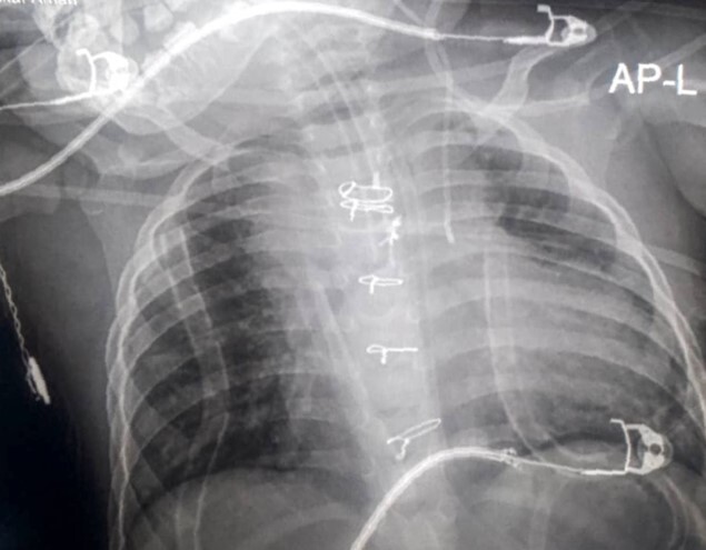
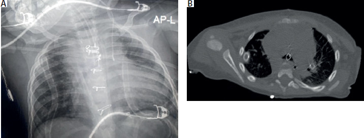
The child was then transferred to the radiology unit for imaging. Correct ET placement with the tip at the carina was demonstrated on a plain chest radiograph (Figure 1A). Computed tomography (CT) did not reveal clear evidence of tracheobronchial pathology; however, it confirmed correct nasogastric tube position and verified the absence of fibrosis, adhesion, and suture fixation (Figure 1B).
FIGURE 1
A) Plain chest X-ray showing endotracheal tube (ET) at carina. B) Axial CT image, which establishes ET position with no airway abnormality
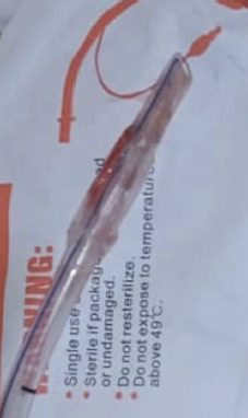
The child was transferred back to the ICU, where a stepwise extubation protocol was initiated. Sedation with ketamine 5 mg, propofol 10 mg, and midazolam 1 mg, together with glycopyrrolate 0.1 mg, was administered. Continuous oxygenation was provided via Ayre’s T-piece. Under direct laryngoscopy, sequential cuff inflation–deflation and careful 180° ET rotation were performed, followed by gentle and sustained spiral traction. The ET immobility gradually resolved with continued traction. Eventually, successful release and removal of the ET were achieved. The removed ET demonstrated a narrowed distal lumen obstructed by a mucus plug and a ring-like depression on the collapsed cuff (Figure 2). After extubation, stable oxygenation and haemodynamics were maintained. There was no airway oedema, bleeding, or stridor. The postoperative course was uneventful, and the child was discharged on day 5.
FIGURE 2
Extubated endotracheal tube showing distal constriction with mucus plug and circumferential cuff anomaly
A stuck ET is an uncommon yet potentially life-threatening airway complication. A large-scale study of 137,866 tracheal intubations reported a difficult extubation incidence of 0.06–0.1%, with stuck ET comprising a considerable proportion [4]. Although rare, a stuck ET may lead to complete airway obstruction, tracheal laceration, subcutaneous emphysema, pneumothorax, or cardiopulmonary collapse if poorly managed [5, 6].
In our patient, despite adherence to standard airway management protocols, extubation was impeded, possibly by distal ET fixation. Ultimately, the ET was removed through systematic measures, emphasising patience, direct visualisation, imaging, and multidisciplinary care. The presence of a mucus plug and circumferential cuff deformity suggested mechanical obstruction compounded by cuff deformation, theorised to result from prolonged intubation, excessive cuff volume and pressure, or inadequate humidification during mechanical ventilation. These mechanisms are inferred rather than directly demonstrated [1, 2, 7].
Similar scenarios with varying aetiologies and management strategies have been described. Panda et al. [8] reported a stuck ET resulting from cuff folding, relieved by cuff re-inflation. Bamgbade et al. [9] observed cuff herniation managed via circumrotation and topical lidocaine. Adhikari et al. [10] described a stuck ET caused by airway oedema and granulation following prolonged intubation, resolved with gentle traction. Probert et al. [11] employed a cricoid split to manage subglottic fixation, and Silva et al. [12] successfully removed a stuck ET with an over-inflated cuff using laryngeal scissors. Suman et al. [13] documented impaired cuff deflation caused by pilot balloon occlusion from tight taping, resolved by obstruction release. Cornforth [14] reported cuffdeflation failure due to a faulty pilot valve, corrected by cutting the pilot tubing. Yau et al. [15] managed a nondeflating cuff by needle puncture, and Tashayod et al. [16] removed a stuck ET using a controlled 180° rotational manoeuvre. Collectively, these cases reinforce the diverse aetiologies of stuck ET and underscore the value of a cautious, systematic approach comprising glottis visualisation and imaging.
Traumatic tracheal intubation may result in a stuck ET secondary to airway injury, with subsequent fibrosis, granulation, and adhesions. In our case, a complete airway assessment was performed. The ET was verified to be appropriate for age, the pilot balloon was tested, and the tube was lubricated. Anaesthesia and airway management were conducted by an experienced clinician [1, 2].
The DAS guidelines state that tracheal extubation is an elective procedure and outline four key steps: plan, prepare, perform, and provide postextubation care [2]. Planning should exclude airway trauma and all causes of airway obstruction, with a clear reintubation plan. Preparation includes complete neuromuscular blockade reversal, adequate suctioning, and cuff deflation. Tracheal extubation should be performed in a controlled setting with vigilant and comprehensive postextubation care [2, 5].
Strict adherence to airway management guidelines produces favourable results. A high index of suspicion is essential. The anaesthesia provider should be unhurried, methodical, and systematic. Direct or video laryngoscopy allows continuous and dynamic visualisation of the pharynx, larynx, and trachea. Plain radiography and CT imaging, combined with radiologist review, ruled out structural anomalies [1, 2].
Forceful traction leading to tracheal laceration, blind cuff puncture, or pilot-tube cutting are potential pitfalls that were avoided. Evidencebased sequential cuff inflation and deflation alongside 180° ET rotation facilitated release of the ET [2].
Safety should be prioritised. Continuous oxygenation, monitoring, spontaneous ventilation, and stable haemodynamics should be maintained during extubation attempts. Early recognition of airway complications is crucial, necessitating immediate cessation of the procedure. Agitation, straining, and coughing should be avoided to allow accurate airway evaluation [1, 2].
Prophylactic corticosteroids can reduce airway oedema and adhesion formation and are useful during prolonged intubation. Video laryngoscopy or flexible bronchoscopy enables precise localisation of the point of resistance. Any intervention should be methodical, structured, and patientcentred. Complete cuff deflation, firm, sustained, and gentle traction under direct or video laryngoscopy, along with repeated inflation–deflation cycles to release folds and adhesions, are beneficial measures. If persistent resistance is encountered, further imaging and specialist review are warranted. Re-intubation equipment and/or surgical airway devices should be prepared during any re-attempt, in case of airway collapse or failed extubation [5].
An individualised approach to care is advised. This structured, evidencebased protocol supports safe and effective management and can ensure successful outcomes even in lowresource settings lacking advanced equipment and immediate otorhinolaryngology support [1–3].
A stuck ET is a rare but life-threatening airway complication. Its safe management demands evidencebased proactive preparedness and multidisciplinary care. Adherence to standardised extubation protocols promotes positive outcomes and ensures patient safety.