Dear Editor,
In recent years, there has been a concerning trend of increased production and abuse of new psychoactive substances, known as “legal highs.” One of the most popular groups includes compounds with psychostimulant effects, such as synthetic cathinones, which are structural analogs of cathinone (S-(-)-2-amino-1-phenylpropan-1-one). Based on their strength as inhibitors of the dopamine transporter, norepinephrine transporter, and serotonin transporter, as well as their ability to release neurotransmitters, they have been divided into three groups:
Cathinones with effects similar to cocaine and 3,4-methylenedioxymeth-amphetamine, e.g., mephedrone.
Methamphetamine-like cathinones, e.g., methylcathinone.
Pyrovalerone cathinones, e.g., α-pyrrolidinopentiophenone (α-PVP).
The consumption of synthetic cathinones can induce effects that are dangerous to the health and life of the potential user. These effects include, among others: tachycardia, hypertension, myocarditis, cardiac arrest, irritability, aggression, panic attacks, seizures, dilated pupils, paresthesia, disseminated intravascular coagulation (DIC), and serotonin syndrome [1].
Serotonin syndrome (serotonergic) is a potentially life-threatening condition caused by excessive stimulation of serotonin receptors. This syndrome can be induced by taking two drugs from the group of serotonin reuptake inhibitors (SSRIs) or monoamine oxidase inhibitors (MAO inhibitors). Diagnosis of serotonin syndrome requires meeting at least two symptoms from the Hunter criteria (Table 1). The prevalence of serotonin syndrome is not fully determined, due to the diverse symptoms of this condition [2]. Serotonin syndrome can be mild or severe. Iatrogenic forms are usually mild, while those induced by psycho-active substances can be severe and sometimes lead to death. This condition usually requires treatment in an intensive care unit (ICU) [2–4].
TABLE 1
Hunter serotonin toxicity criteria [4]
According to literature data, the effects of synthetic cathinones can appear within 15–45 minutes after ingestion and last from 2 to 7 hours. However, adverse effects can persist from several hours to several days [5, 6].
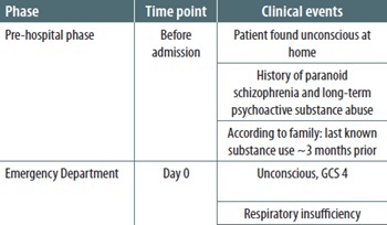
A 24-year-old male patient with a history of paranoid schizophrenia was found unconscious at home. According to the patient’s family, the last known use of psychoactive substances had occurred approximately three months prior to admission. The patient had a long history of psychoactive substance abuse and had undergone multiple detoxification treatments. His regular medications included olanzapine 20 mg per day and levomepromazine 25 mg per day.
The patient was transported by Emergency Medical Services to the Emergency Department. Upon admission, he was unconscious and in severe respiratory failure. Neurological examination revealed narrow, symmetrical, light-reactive pupils. The patient scored 4 points on the Glasgow Coma Scale (GCS). Physical examination demonstrated a soft abdomen without signs of peritoneal irritation. Rectal examination did not reveal evidence of lower gastrointestinal bleeding.
Due to respiratory failure and impaired consciousness, the patient was intubated and mechanically ventilated. Blood pressure on admission was 100/50 mmHg, and marked tachycardia of 170 beats min–1 was observed. Chest radiography excluded pneumothorax and cardiac tamponade. Noradrenaline infusion was initiated. Activated charcoal was administered through a gastric tube (60 tablets).
Initial laboratory investigations demonstrated markedly elevated troponin levels (367.70 ng L–1), acute liver and kidney injury (ALT 279 U L–1,
AST 400 U L–1, eGFR 22.61 mL min–1 1.73 m–2, anuria), leukocytosis (WBC 25.28 × 10³ μL–1), thrombocytopenia (PLT 96 × 103 μL–1), and severe hypofibrinogenemia (fibrinogen < 40 mg dL–1). No signs of anemia were present. Arterial blood gas analysis revealed severe mixed respiratory and metabolic acidosis (pH 6.94, pCO2 95 mmHg, HCO3– 20.4 mmol L–1, standard HCO3– 14.0 mmol L–1).
After initial stabilization in the Emergency Department, the patient was transferred to the Department of Anesthesiology and Intensive Care. During the first hours of hospitalization, the patient developed generalized muscle tremors and symmetrical myoclonus, suggestive of serotonin syndrome [2]. Treatment with midazolam, crystalloids, amiodarone, and b-blockers was initiated. Despite escalating doses of catecholamines, progressive hypotension developed, with blood pressure decreasing to 50/35 mmHg.
Following partial hemodynamic stabilization, a dialysis catheter was inserted into the right femoral vein, and continuous hemodiafiltration was initiated. Toxicological analysis of blood and urine samples qualitatively demonstrated high-intensity signals corresponding to N-ethyl-pentedrone (NEPD). Additionally, pharmacological agents administered during treatment, including amiodarone, lido-caine, and midazolam, were detected.
In the following hours, the patient developed signs of refractory shock with a suspected septic component. Systemic glucocorticoid therapy was initiated. At that time, the computed tomography (CT) scanner in the hospital was out of service. Because of the patient’s rapid hemodynamic deterioration and profound clinical instability, transportation to another hospital for CT was considered impossible. Despite maximal vasopressor support, further circulatory deterioration occurred.
Approximately three hours after ICU admission, the patient no longer responded to fluid resuscitation. Forty-five minutes later, pulseless electrical activity (PEA) cardiac arrest occurred. Cardiopulmonary resuscitation was initiated immediately. Return of spontaneous circulation was temporarily achieved, although severe circulatory insufficiency persisted, with blood pressure measuring 60/15 mmHg. Treatment included further crystal-loid administration, repeated doses of adrenaline and sodium bicarbonate, and continuation of maximum-dose catecholamine infusions.
Despite appropriate mechanical ventilation, progressive hypercapnia persisted. Hemodynamic monitoring demonstrated markedly reduced vascular resistance and cardiac output. Pupils became bilaterally dilated and non-reactive to light. Continuous renal replacement therapy was maintained throughout treatment.
Approximately 30 minutes later, recurrent cardiac arrest in the form of PEA progressing to asystole occurred. Advanced resuscitation efforts were continued, including chest compressions and additional adrenaline administration; however, no effective hemodynamic rhythm was restored. Given the exhaustion of all available therapeutic options, the patient was pronounced dead.
Law enforcement authorities were notified because of suspected third-party involvement, and the patient’s family was informed of the death.
Two weeks later, a medico-legal autopsy was performed. Internal examination demonstrated cerebral edema and congestion of the meninges. The respiratory tract contained dark tarry material corresponding to hemorrhagic gastrointestinal contents extending from the esophagus to the small intestine. Pulmonary edema was also observed. Histopathological examination of the stomach revealed inflammatory infiltrates, hemorrhagic lesions, and erosions of the gastric mucosa. Hepatic steatosis was additionally identified.
Postmortem toxicological analysis of blood and urine samples using liquid chromatography–mass spectrometry (LC/MS) qualitatively confirmed the presence of NEPD. The analysis additionally detected medications administered during hospitalization, including amiodarone, lidocaine, midazolam, and a-hydroxymidazolam. Quantitative LC-MS/MS analysis demonstrated NEPD concentrations of 566 ng mL–1 in blood and 9725 ng mL–1 in urine.
Ultimately, the cause of death was determined to be massive gastrointestinal hemorrhage associated with hemorrhagic gastropathy and severe multi-organ failure following NEPD intoxication.
Table 2 provides a chronological overview of key clinical events.
TABLE 2
Timeline of clinical course
Blood and urine samples were also collected for toxicological analysis. The analysis was conducted for non-volatile organic compounds using LC-MS. To 100 μL of whole blood and 100 μL of urine, 10 μL of internal standard diazepam D5 (concentration 500 ng mL–1) was added, deproteinized with 300 μL of acetonitrile, mixed, centrifuged, and 100 μL of supernatant was diluted with 500 μL of deionized water for analysis. Separation was performed using a Shimadzu Nexera XR apparatus (Kyoto, Japan) and an AB Sciex Triple-TOF 5600+ mass spectrometer (Framingham, MA, USA), with a DuoSpray ion source (APCI/ESI) and Calibrator Delivery System (CDS). The stationary phase consisted of a monolithic Chromolithic Performance RP-18e, 2–100 mm column (Merck, Darmstadt, Germany). The mobile phase consisted of two solutions: (A) 0.1% formic acid in water and (B) 0.1% formic acid in acetonitrile. The analysis was carried out at a flow rate of 0.4 mL min–1 with a gradient slope from 95% to 5% (A) over 10 minutes. Data analysis was performed using Peak View v. 2.1 2 Master View software.
Subsequently, a quantitative analysis of NEPD was performed. To 200 µL of blood and 200 µL of urine, 10 µL of internal standard in the form of a-PVP D8 at a concentration of 10 µg mL–1 and 200 µL of 0.5 M ammonium carbonate solution (pH 9) were added, and twice extracted with 1 mL of ethyl acetate. After extraction and centrifugation, the organic phase was evaporated to dryness in a vacuum centrifuge at 40ºC. The dry residue was dissolved in 100 µL of mobile phase mixture 90% (A) and 10% (B). The prepared samples were analyzed by LC-MS/MS using an Agilent 1200 series liquid chromatograph (Agilent, USA) and a Poroshell 120 EC C18 3.0 × 75 mm, 2.7 µm chromatographic column (Agilent, USA). Phase A was 0.1% formic acid in water, and phase B was 0.1% formic acid in acetonitrile, in a gradient mode (Table 3).
TABLE 3
Gradient mode
| Time [min] | Flow [µl min–1] | Phase A [%] | Phase B [%] |
|---|---|---|---|
| 0 | 500 | 90.0 | 10.0 |
| 1.00 | 500 | 90.0 | 10.0 |
| 7.00 | 500 | 60.0 | 40.0 |
| 9.00 | 500 | 60.0 | 40.0 |
| 10.00 | 500 | 90.0 | 10.0 |
Mass spectrometer: Agilent 6410B (Agilent, USA), ion source: electro-spray, positive ionization, gas temperature 300ºC, gas flow 8 L min–1, nebulizer 40 psi, capillary 4000 V. Scanning mode: MRM with ion transitions for each analyzed compound (Table 4).
TABLE 4
Analysis of individual compounds in mass spectrometry
| Name | Q1 Mass [Da] | Q3 Mass [Da] | Fragmentor [V] | CE (volts) |
|---|---|---|---|---|
| N-ethyl-pentedrone 1 | 206.2 | 130.1 | 90 | 29 |
| N-ethyl-pentedrone 2 | 206.2 | 91.1 | 90 | 25 |
| α-PVP D8 | 240.2 | 91.1 | 130 | 21 |
| α-PVP D8 | 240.2 | 77.1 | 130 | 45 |
The calibration curve for NEPD (Cayman Chemical, USA) was established using prepared standard solutions diluted in a matrix-matched solution. Calibration curve range: 10–500 ng mL–1, R2 = 0.998. In order to evaluate the dilution integrity, plasma QC sample at concentration above the ULOQ was diluted by 10 and 100% with the blank plasma. In the prepared plasma replicates (n = 5) the analytes were determined with accuracy of 94.4% and precision (CV) of 3.5%. Thus, it was proved that the developed HPLC-MS/MS method can be applied to analyze plasma samples diluted due to the analyte concentration exceeding the ULOQ. Data analysis was performed using MassHunter Workstation software (ver. B 08.00).
N-ethyl-pentedrone (2-(ethylamino)-1-phenyl-1-pentan-1-one) is a synthetic cathinone belonging to the second generation of cathinone derivatives that emerged on the recreational drug market in the mid-2010s [7]. Similar to other synthetic cathinones, NEPD primarily affects monoaminergic neurotransmission through interactions with dopaminergic, noradrenergic, and serotonergic transporters [8]. The most common routes of administration include intra-nasal use and vaporization, although the route of administration mainly influences the onset of effects rather than the overall toxicity of the compound [9].
In the present case, a 24-year-old patient died following severe NEPD intoxication. On admission, the patient was unconscious (GCS 4) and developed generalized tremors and symmetrical myoclonus during the early phase of hospitalization, which are characteristic manifestations of serotonin syndrome [2]. In addition, the patient rapidly developed signs of multi-organ failure, including acute toxic injury to the liver and kidneys, severe metabolic acidosis, circulatory instability, and progressive shock. Despite treatment in the intensive care unit, including vasopressor therapy, mechanical ventilation, and continuous renal replacement therapy, the patient’s condition progressively deteriorated, ultimately resulting in circulatory collapse and cardiac arrest.
The diagnosis of serotonin syndrome remains primarily clinical and is most commonly based on the Hunter Serotonin Toxicity Criteria, which demonstrate high sensitivity and specificity for serotonin toxicity [4]. The Hunter criteria require exposure to a serotonergic agent together with characteristic neuromuscular and autonomic manifestations, including spontaneous or inducible clonus, tremor, hyperreflexia, agitation, hyperthermia, or ocular clonus [3, 4]. In the present case, the occurrence of tremors and myoclonus in the setting of confirmed NEPD exposure strongly supports serotonin toxicity as an important component of the clinical presentation.
Toxicological analysis confirmed the presence of NEPD at concentrations of 566 ng/mL in blood and 9725 ng mL–1 in urine. These concentrations were markedly higher than toxic levels previously reported in the literature. Pieprzyca et al. [10] reported toxic blood concentrations ranging from 7 to 137 ng mL–1, with a mean concentration of 44 ng mL–1 and a median concentration of 18 ng mL–1. Importantly, in the present case, blood samples were collected post mortem after several hours of hospitalization and intensive medical interventions, suggesting that the peak concentration during life may have been even higher. Therefore, the extremely high concentration detected in this patient strongly supports a causal relationship between NEPD intoxication and the fatal outcome.
Interpretation of postmortem concentrations of synthetic cathinones should be performed cautiously because these compounds may undergo postmortem redistribution and degradation in biological material [11]. Synthetic cathinones demonstrate low to moderate postmortem redistribution; however, their stability is strongly dependent on pH conditions [12–14]. Acidic environments tend to limit degradation, whereas alkaline conditions may significantly accelerate decomposition [10, 12, 13]. Additionally, postmortem biochemical changes occurring during sample storage may further influence measured concentrations. Consequently, measured postmortem NEPD levels may underestimate concentrations present during life.
Autopsy findings revealed massive gastrointestinal hemorrhage as the immediate cause of death in the setting of severe multi-organ failure. Laboratory investigations demonstrated thrombocytopenia, severe hypofibrinogenemia, leukocytosis, and markedly elevated procalcitonin levels. Although complete coagulation parameters, including D-dimer concentrations, were unavailable, the overall clinical and laboratory picture strongly suggested disseminated intravascular coagulation (DIC), most likely associated with severe systemic inflammation and shock [3].
Several mechanisms may explain the hemorrhagic complications observed in this patient. Synthetic cathinones inhibit serotonin reuptake similarly to amphetamine derivatives [8]. Platelets are unable to synthesize serotonin independently and rely on serotonin uptake from plasma. Consequently, inhibition of serotonin transport impairs platelet aggregation and hemostasis, thereby increasing the risk of bleeding [2]. This mechanism may partially explain the extensive gastrointestinal hemorrhage observed in the present case.
Additional contributing factors likely included prolonged shock and glucocorticoid therapy. Severe shock results in impaired gastrointestinal perfusion and mucosal ischemia, pre-disposing patients to erosions and ulceration [3]. Simultaneously, glucocorticoids reduce prostaglandin synthesis, impair mucosal protective mechanisms, and increase susceptibility to gastrointestinal bleeding [3]. The coexistence of serotonin-related platelet dysfunction, circulatory failure, and glucocorticoid therapy likely contributed significantly to the development of fatal hemorrhagic gastropathy.
Synthetic cathinones have been associated with numerous severe systemic complications, including rhabdomyolysis, renal failure, metabolic acidosis, cardiovascular instability, hepatic injury, and coagulopathy [8, 15, 16]. Such complications may pre-dispose patients to progression toward multi-organ failure. Similar findings have previously been described in intoxications involving pentedrone, a structurally related compound differing from NEPD only by substitution of an ethyl group at the nitrogen atom [17]. Rojek et al. [17] described a fatal case of pentedrone intoxication complicated by rhabdomyolysis, acute kidney injury, liver damage, disseminated intravascular coagulation, and massive gastrointestinal bleeding. Comparable clinical and pathological findings were observed in the present patient, including extensive gastrointestinal hemorrhage and toxic injury to the liver and kidneys.
Although serotonin syndrome has rarely been explicitly diagnosed in previously published NEPD intoxication cases, many reported clinical manifestations overlap with classical features of serotonin toxicity [15, 18]. Experimental studies additionally demonstrated that synthetic cathinones may induce hyperthermia, aggression, behavioral disturbances, and weight loss through dopaminergic and serotonergic mechanisms [19–22]. These findings further support the concept of profound systemic toxicity associated with NEPD exposure.
Recent pharmacokinetic studies demonstrated that NEPD possesses a relatively long elimination half-life of approximately 770 minutes and undergoes extensive metabolism with formation of multiple metabolites [23]. These pharmacokinetic properties may contribute to prolonged toxic effects and accumulation within the body, particularly following high-dose exposure.
The present case demonstrates a fulminant course of NEPD intoxication characterized by severe serotonin toxicity, refractory shock, multi-organ failure, coagulopathy, and fatal gastrointestinal bleeding. Given the increasing prevalence of synthetic cathinones and the still limited toxicological data concerning NEPD, clinicians should remain vigilant for severe complications, particularly in patients presenting with neuromuscular abnormalities, organ dysfunction, circulatory instability, or signs of coagulopathy [8, 11, 15, 24].
Further clinical observations and additional well-documented case reports are necessary to better characterize the toxicological profile of NEPD, establish reliable toxic and lethal concentration ranges, and improve management strategies for patients exposed to this compound.
In the present case, fatal intoxication with NEPD was associated with very high blood concentrations of the substance and a rapidly progressive clinical course leading to death within hours of admission. The presence of tremor and myoclonus, together with rapid deterioration and multi-organ failure, indicates that serotonin syndrome was a significant component of the clinical presentation.
The coexistence of hypofibrinogenemia, thrombocytopenia, leukocytosis, and markedly elevated procalcitonin levels strongly suggests the development of disseminated intravascular coagulation, likely of septic origin, contributing to massive gastrointestinal bleeding identified at autopsy.
This case demonstrates that NEPD intoxication may lead to a combination of serotonin toxicity, multi-organ failure, and coagulopathy, resulting in a fulminant and fatal outcome.
We confirm that formal permission from the Prosecutor’s Office was obtained to use the collected material for scientific and research purposes. Additionally, the case report does not include any identifiable personal data, and all sensitive information has been anonymized in accordance with medical confidentiality requirements and applicable regulations.