


Dear Editor,
Perioperative management of anti-thrombotic therapy is a major concern in high-risk patients. Emergency cases can be even more challenging to address, due to limited timeframes. We report a case of a post-ST-elevation myocardial infarction (STEMI) patient on dual antiplatelet therapy requiring emergency laparotomy. Written informed consent for publication was obtained from the patient.
A 54-year-old man was admitted to the emergency department with symptoms consistent with myocardial infarction. His medical history was significant for arterial hypertension, non-insulin dependent diabetes mellitus, dyslipidaemia, smoking, daily alcohol consumption and obesity class II (body mass index 39.2 kg m–2). He had previously undergone plastic mesh reconstruction of incisional hernia twice.
Based on clinical and laboratory findings, the patient was diagnosed with inferior STEMI with right ventricle involvement. He was transferred to the catheterization laboratory for a coronary angiography, where he was diagnosed with critical right coronary artery stenosis and placed on three stents (Promus Premier 3.00 × 24 mm, 3.00 × 20 mm and 3.5 × 12 mm). The patient was then transferred to the cardiac Intensive Care Unit (ICU) for observation. A loading dose of 60 mg of prasugrel was administered followed by 10 mg daily in combination with 100 mg of acetylsalicylic acid daily. Other medical treatment included bisoprolol, rosuvastatin, empagliflozin, vildagliptin/metformin hydrochloride and omeprazole.
Five days after the percutaneous coronary intervention (PCI), while still in the hospital, the patient experienced acute pain in the right lateral abdominal area. Laboratory tests revealed an elevated white blood cell (WBC) count (22,940 μ L–1, normal range 4000– 11,000 μL–1) and a C-reactive protein (CRP) level of 146 mg L–1 (normal level < 3 mg L–1). Computed tomography showed an enlarged, ruptured appendix along with free fluid in the right iliac fossa. Because of the high perioperative risk, the patient was initially administered intravenously conservative treatment including ceftriaxone, metronidazole and amikacin. However, both laboratory results (WBC 25,430 μL–1, CRP 342 mg L–1) and the patient’s clinical condition deteriorated over the following hours, requiring emergency surgery.
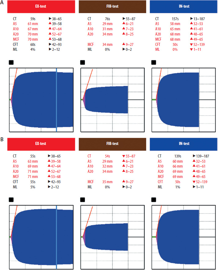
When the patient was deemed eligible for surgery, 24 h had elapsed since the last dose of prasugrel, which increased the risk of intraoperative haemorrhage. Blood samples were obtained just prior to surgery for viscoelastic tests and also a platelet aggregation study. Platelets had also been ordered and prepared for potential transfusion in the hospital blood bank. ClotPro (Enicor GmbH, Munich) EX-, IN- and FIB-test results (evaluating the extrinsic, intrinsic pathway and fibrinogen function respectively) were readily available and did not indicate any impairment of the patient’s haemostatic pathways (Figure 1A), which initially raised the concern that prasugrel may not have been an appropriate antiplatelet for this patient.
FIGURE 1
Thromboelastometry results (ClotPro, Enicor GmbH, Munich) (A) 30 min preoperatively, (B) 6 h postoperatively
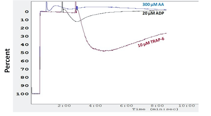
Aggregometry results became available 3 h after venipuncture. Three different agonists were used: 300 μM ara chidonic acid (AA, A3611, Sigma- Aldrich, St. Louis, MO, USA) and 10–20 μM adenosine diphosphate (ADP, PPR-P 384, Chrono-Log, Havertown, PA, USA). The light transmission aggregation (LTA) method was applied. No platelet aggregation was observed in response to AA (500 μM) or ADP (10 or 20 μM), with < 10% aggregation. In contrast, thrombin receptor activating peptide 6 (10 μM) induced 51 ± 2% aggregation. These results indicate sustained PAR-1 activation and a significant reduction in COX-1 and P2Y12 aggregation. Pharmacological inhibition of platelets, e.g. dual antiplatelet therapy (aspirin and P2Y12 inhibitor), is most consistent with this. Clinically, it indicates an increased risk of perioperative bleeding (Figure 2).
FIGURE 2
Representative curve of platelet aggregation induced by adenosine diphosphate (20 μΜ) (black curve), arachidonic acid (300 μΜ) (blue curve) and thrombin receptor activating peptide 6 (10 μΜ) (red curve). The X-axis represents the aggregation time and the Y-axis represents the percentage of platelet aggregation
Based on this information and with no evidence of bleeding at the surgical site, the platelet transfusion was withheld. A midline incision was performed. Exploratory laparotomy revealed generalised purulent peritonitis as a result of a ruptured appendix. An arduous appendectomy, peritoneal lavage and diligent haemostasis was performed. The operation lasted for 70 min and was uneventful. After recovering from anaesthesia, the patient was successfully extubated and returned to the cardiac ICU. A second set of viscoelastic tests was run 6 h postoperatively, and all values were within normal limits (Figure 1B).
Total blood loss in the vacuum drain was 20 mL during the first 24 h postoperatively, and the patient was started again on the prior dual antiplatelet therapy on postoperative day 1 (POD-1). The rest of his postoperative hospital stay was uneventful, and he was discharged on postoperative day 10 (POD-10). At 30 days postoperatively, the patient had no major complications associated with the surgery or the PCI.
Dual antiplatelet therapy is recommended for 12 months following PCI for STEMI, unless contraindications exist [1]. Perioperative antiplatelet management is often challenging, as both the thrombotic risk of the patient and the haemorrhagic risk of the procedure must be considered. In general, a preoperative delay of 5–7 days is recommended for prasugrel prior to major surgical procedures with or without bridging with cangrelor [2]. None of these options were viable, one because of a recent stent placement and the other because of the urgency of the surgery. According to the literature, dual antiplatelet therapy must not be interrupted in the case of emergency surgery so close to PCI, despite the increased possibility of bleed- ing [3]. Urgent surgery on an antiplatelet-therapy patient may cause stress to the medical team and dictate unnecessary platelet transfusions in the absence of point-of-care tests to access haemostasis.
LTA remains the gold standard method for evaluating platelet function but has the drawback that it is not widely available in practice, and it is time-consuming. On the other hand, visco-elastic testing is a fast and easy-to-use method for assessing the patient’s clotting status and offers various methods for assessing different aspects of haemostasis. The combination of both techniques allowed for a personalized assessment of the patient’s clotting status, confirming that although antiplatelet therapy was effective in preventing platelet aggregation and therefore clot formation, the remaining coagulation mechanisms were sufficient to prevent major perioperative bleeding.
Despite the fact that platelet aggregation induced by AA and ADP was inhibited by their respective anti-platelet agents, clot formation was still possible because the thrombin pathway was not inhibited. To inhibit the thrombin pathway, specific thrombin inhibitors should be prescribed to patients. However, thrombin inhibitors are not part of routine antiplatelet therapy, as the efficacy and safety of such an add-on has not been fully established [4].
In pursuing the optimal antithrombotic balance – the “sweet spot” in anti-thrombotic therapy – this case may suggest that the current guidelines for withholding antithrombotic therapy may be too restrictive and should be followed by shorter or no interruption of treatment in patients with otherwise intact coagulation mechanisms undergoing low bleeding risk procedures.

