Thyroid hormone dysregulation is a hallmark of critical illness and is known as non-thyroidal illness syndrome (NTIS) or euthyroid sick syndrome. The presence of low triiodothyronine and/or thyro-xine concentrations with inappropriately normal or low thyroid-stimulating hormone (TSH) suggests a profound alteration of endocrine homeostasis in response to systemic stress [1–3]. NTIS is not a primary form of thyroid disorder, but is an adaptive (or possibly maladaptive) response to a severe illness, such as sepsis, trauma, and major surgery [3–5].
Importantly, the severity of thyroid hormone disturbances is associated with disease burden and has been shown to be associated with worse outcomes and increased mortality in critically ill patients [2, 3]. Yet, whether these changes are protective or modifiable therapeutic targets remains unclear. Despite a strong biological rationale, clinical evidence supporting thyroid hormone supplementation in this context is limited, heterogeneous, and largely based on small or pilot studies, with no clear guidance for clinical practice [6].
There has been little real-world evidence on the biochemical and clinical effects of thyroid hormone supplementation in unselected ICU populations. Also, it is uncertain whether the change in thyroid hormone concentrations after thyroid hormone supplementation is due to true therapeutic effects or to the severity of the underlying disease. Therefore, the present study aimed to assess thyroid hormone profiles and the effects of thyroid hormone supplementation in critically ill patients, particularly in terms of their association with disease severity and clinical outcomes.
Methods
This study was conducted as a retrospective, observational, single-center analysis carried out in Gdańsk, Poland. The study protocol received approval from the Bioethics Committee for Scientific Research at the at the Medical University of Gdańsk. Clinical records of patients admitted to the Intensive Care Unit of the Intensive Care Unit of the University Clinical Center (UCK) in Gdańsk between 2023 and 2024 were reviewed and analyzed. Due to retrospective character of the study, the Bioethics Committee for Scientific Research waived the need for informed consent.
Patients
All eligible patients admitted to the Intensive Care Unit (ICU) at the University Clinical Center (UCK) in Gdańsk between 2023 and 2024 were included in this retrospective analysis. Collected clinical data comprised demographic characteristics, indications for ICU admission, details of the ICU course, and treatment outcomes. In addition, information related to thyroid hormone supplementation (including dosage, timing, and route of administration) was evaluated, together with laboratory measurements of thyroid function such as TSH, free triiodothyronine (fT3), and free thyroxine (fT4) concentrations. Given the retrospective and observational nature of the study, the institutional review board waived the requirement for obtaining informed consent from patients. The inclusion criteria were: (i) ICU hospitalization between 2023 and 2024, and (ii) receipt of thyroid hormone supplementation during the ICU stay. All patients received ICU management in accordance with current medical guidelines under the supervision of a specialist in anesthesiology and intensive therapy. In the absence of universally accepted clinical guidelines within the field of thyroid hormone supplementation in the ICU, the decision to initiate thyroid hormone supplementation was made empirically by the treating intensivist and was typically prompted by a combination of biochemical findings (most commonly low fT3 and/or fT4 with inappropriately non-elevated TSH) and clinical context (disease severity and the absence of clinical improvement). The choice between levothyroxine and liothyronine, as well as the specific dosing regimen, was based on individual clinical judgment, disease severity, and the specific pattern of biochemical abnormalities. Shock was defined as persistent arterial hypotension (mean arterial pressure < 65 mmHg) despite adequate fluid resuscitation, requiring vasopressor therapy to maintain target perfusion pressure [7]. Acute kidney injury was diagnosed according to KDIGO [8].
Statistical analysis
Categorical variables are presented as counts (%). Normality of continuous variables was assessed using the Shapiro-Wilk test and inspection of Q–Q plots. Normally distributed data are expressed as mean ± standard deviation (SD), and non-normally distributed data are expressed as median [IQR]. Values below the lower limit of quantification (LOQ) were imputed as LOQ/2. Group comparisons were performed using the c2 test or Fisher’s exact test for categorical variables and the Mann-Whitney U test for continuous variables. Correlations were assessed using Spearman’s rank correlation coefficient. To account for multiple testing, P-values were adjusted with the Benjamini-Yekutieli procedure. Relative changes were calculated as Δ = (subsequent value − initial value)/initial value × 100%. All tests were two-sided, and P < 0.05 was considered statistically significant. Statistical analyses were performed using GraphPad Prism (GraphPad Software, USA). Paired first-versus-last comparisons were performed using the Wilcoxon signed-rank test. Longitudinal thyroid hormone changes and correlation heatmaps were generated using Python 3.14.2 with pandas, Matplotlib, SciPy, and seaborn.
Results
Study population and baseline characteristics
The study population consisted of 98 critically ill patients, of whom 51% were female. The median age was 69 years [IQR 49–76], with a median body mass index (BMI) of 27.83 kg m–2 [IQR 23.31–32.64]. The most common reasons for ICU admission were respiratory failure (26%), neurological conditions (21%), and circulatory failure (20%). The predominant comorbidities included hypertension (51%), diabetes mellitus (24%), and chronic kidney disease (17%). Disease severity was reflected by median APACHE II, SAPS II, and SOFA scores of 17 [IQR 11–22], 40 [IQR 29–50], and 9 [IQR 7–11], respec-tively. The median ICU length of stay was 14 days [IQR 9–20], with mechanical ventilation and vasopressor support required for a median of 10 days each. ICU mortality was 29%, and a futile therapy protocol was applied in 10% of patients (Table 1).
TABLE 1
Demographic and clinical characteristics of patients (N = 98)
Thyroid hormone profiles and preexisting thyroid disease
Patients with and without preexisting thyroid disease did not differ significantly in disease severity, with comparable SOFA (6 [IQR 3–8] vs. 7 [IQR 2–9]; P = 0.304), APACHE II (15 [IQR 10–22] vs. 18 [IQR 12–22]; P = 0.322), and SAPS II scores (40 [IQR 27–51] vs. 40 [IQR 33–50]; P = 0.993). Lactate concentrations were non-significantly higher in patients with pre-existing thyroid disease than in those without preexisting thyroid disease (1.4 vs. 1.0 mmol L–1; P = 0.066). TSH and fT3 were also similar between groups (P = 0.280 and P = 0.112, respectively), whereas fT4 was significantly higher in patients with preexisting thyroid disease (11.56 [IQR 7.90–14.15] vs. 8.46 [IQR 7.36–10.70] pmol L–1; P = 0.010). Regarding thyroid hormone supplementation, patients with preexisting thyroid disease received lower doses of liothyronine (0 [IQR 0.0–7.5] vs. 7.5 [IQR 7.5–15.0] µg; P < 0.001) and higher doses of levothyroxine (75 [IQR 50.0–118.5] vs. 75.0 [IQR 37.5–75.0] µg; P = 0.002). All patients received thyroid hormone supplementation via the gastrointestinal tract (Table 2).
TABLE 2
Comparison of clinical characteristics between patients with and without preexisting thyroid disease at study inclusion
The distribution of thyroid hormone measurements differed significantly between groups only for TSH (P = 0.003). Patients with preexisting thyroid disease more frequently presented with elevated TSH concentrations (> 4.94 µU mL–1; 37.8% vs. 14.7%), while suppressed TSH (< 0.35 µU mL–1) was more common in those without thyroid comorbidities (21.2% vs. 11.3%); values within the reference range were predominant in both groups (50.9% vs. 64.1%). Regarding fT3, the majority of measurements fell below the lower reference limit (< 1.78 pmol L–1) in both groups (53.7% vs. 57.6%), with no values exceeding the upper limit, and the between-group difference was not significant (P = 0.063). Similarly, fT4 distributions were comparable between patients with and without preexisting thyroid disease (P = 0.892), with approximately half of all measurements falling within the reference range in both groups and none exceeding the upper limit of 19.05 pmol L–1 (Table 3).
TABLE 3
Distribution of measurements according to thyroid-stimulating hormone (TSH), free triiodothyronine (fT3), and free thyroxine (fT4) concentrations relative to laboratory reference ranges
Impact of organ dysfunction and ICU interventions
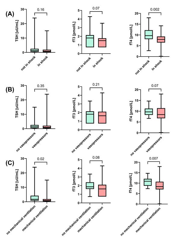
All analyses were performed using the first available laboratory measurements obtained. Baseline TSH concentrations did not differ significantly between patients with and without shock (P = 0.16) or between those requiring and not requiring vasopressor support (P = 0.35). However, patients receiving mechanical ventilation had significantly lower TSH concentrations compared to non-ventilated indivi-duals (P = 0.02). No statistically significant diffe-rences in baseline fT3 concentrations were observed across any of the clinical strata analyzed. Nevertheless, fT3 concentrations were lower in patients with shock than in those without shock (P = 0.07) and in those undergoing mechanical ventilation than in non-ventilated patients (P = 0.08), although these diffe-rences were not statistically significant, whereas no difference was found in relation to vasopressor use (P = 0.21). In contrast, baseline fT4 concentrations were consistently and significantly lower in patients with more severe clinical status. Specifically, reduced fT4 concentrations were observed in patients with shock (P = 0.002) and those receiving mechanical ventilation (P = 0.007). Overall, when considering only the first obtained laboratory values, fT4 demonstrated the most consistent association with markers of disease severity, while TSH and fT3 showed limited or no significant differences across groups (Figure 1).
FIGURE 1
Comparison of first obtained TSH, fT3 and fT4 concentrations between patient groups stratified by clinical status: patients without shock vs. with shock (A), patients not requiring vs. requiring vasopressor support (B), and patients not receiving vs. receiving mechanical ventilation (C). Green bars indicate patients without the respective condition, whereas red bars indicate patients with the condition
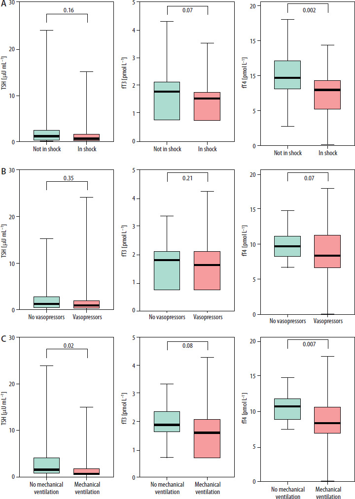
Baseline TSH concentrations did not differ significantly between patients with and without acute kidney injury (AKI) (P = 0.38), between those requiring and not requiring continuous renal replacement therapy (CRRT) (P = 0.75), or between patients exposed and not exposed to iodinated contrast within 3 days prior to hormone assessment (P = 0.28). In contrast, baseline fT3 concentrations were significantly lower in patients with AKI compared to those without AKI (P = 0.01). No significant differences were observed between patients with and without CRRT (P = 0.53) or according to recent iodinated contrast exposure (P = 0.86). Baseline fT4 concentrations were significantly lower in patients with AKI (P = 0.007) and in those requiring CRRT (P = 0.007). In contrast, no significant difference in fT4 concentrations was observed with respect to recent iodinated contrast exposure (P = 0.652) (Figure 2).
FIGURE 2
Comparison of first obtained TSH, fT3, and fT4 concentrations between patient groups stratified by renal and exposure-related clinical status: patients without acute kidney injury vs. with acute kidney injury (A), patients not requiring vs. requiring continuous renal replacement therapy (B), and patients not receiving vs. receiving iodinated contrast within 3 days prior to hormone concentration measurement (C). Green bars indicate patients without the respective condition, whereas red bars indicate patients with the condition
When all available measurements were included in the analysis, several differences compared to the first-value-only approach emerged. For TSH, a significant decrease was observed in patients requiring CRRT, which was not present in the baseline analysis, while other comparisons remained largely unchanged. In contrast, fT3 demonstrated markedly stronger associations, becoming significantly lower across most clinical severity groups (including shock, vasopressor use, mechanical ventilation, AKI, and contrast exposure), whereas previously these differences were limited or non-significant. For fT4, the overall pattern remained consistent, with persistently lower concentrations in more severe conditions (Supplementary Figures 1 and 2).
Correlation analysis of thyroid hormones
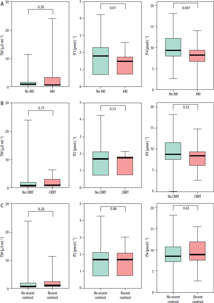
Correlation analysis was performed using Spearman’s rank coefficients with Benjamini-Yekutieli correction for multiple comparisons (Figure 3). When restricted to the first available measurement per patient (panel B), no correlations survived correction, indicating that baseline thyroid hormone values were not robustly associated with the examined clinical variables. In contrast, analysis of all available measurements (panel A) identified several modest but consistent associations: TSH correlated positively with ICU day of sampling (ρ = 0.36, P < 0.01) and levothyroxine dose (ρ = 0.26, P < 0.05), and negatively with SOFA score (ρ = −0.26, P < 0.01). fT3 was also inversely associated with SOFA score (ρ = −0.26, P < 0.01), while fT4 correlated negatively with liothyronine dose (ρ = −0.26, P < 0.05). All effect sizes were small to moderate. Notably, the direction and magnitude of most correlations were similar across both panels, for instance, SOFA–fT3 (ρ = −0.26 vs. −0.34) and liothyronine–fT4 (ρ = −0.26 vs. −0.27), yet none reached significance in the per-patient analysis. This pattern may reflect time-dependent within-patient changes during the ICU stay, though the smaller effective sample size in the per-patient analysis limits statistical power and precludes definitive interpretation. Furthermore, repeated measurements from the same patient introduce within-subject correlation, and hormone concentrations sampled later in the ICU course inevitably coincide with evolving treatment exposure. The observed associations therefore cannot be attributed to any single mechanism.
FIGURE 3
Heatmap of Spearman correlations between thyroid hormone parameters and selected clinical variables. Cell values represent correlation coefficients (Spearman’s ρ), and * indicates significance after Benjamini-Yekutieli adjustment (adjusted P < 0.05). Single-word drug labels refer to drug dose
Temporal changes in hormone concentrations
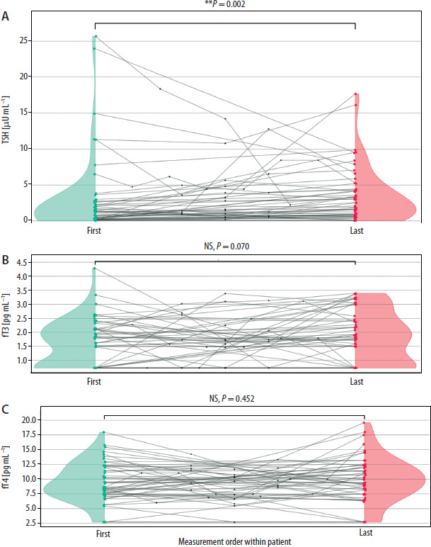
Among patients with repeated hormone measurements (Figure 4), TSH increased significantly between the first and last available measurements (median 1.15 → 2.29 µU mL–1; W = 278.0, P = 0.0025), whereas fT3 (1.61 → 1.77 pmol L–1; W = 245.5, P = 0.07) and fT4 (8.63 → 9.66 pmol L–1; W = 493.0, P = 0.45) did not change significantly. Individual trajectories varied widely, although the majority of patients showed a net increase in TSH between the first and last measurements.
FIGURE 4
Trajectory plots of thyroid hormone concentrations in patients with at least two measurements per hormone (TSH: 52 patients, 138 measurements; fT3: 54 patients, 143 measurements; fT4: 54 patients, 143 measurements). Each line traces one patient’s course, with intermediate values evenly spaced between the first and last measurements. Mint markers and half-violins denote first measurements; salmon markers and half-violins denote last measurements; grey dots indicate intermediate timepoints. First-to-last comparisons were performed using paired Wilcoxon signed-rank tests
Discussion
Thyroid hormone alterations in critically ill patients were primarily associated with disease severity. Lower fT3 and fT4 concentrations were consistently observed in patients with shock, organ support requirements, and higher SOFA scores, while TSH showed less consistent changes. Hormone profiles were largely similar regardless of preexisting thyroid disease. Correlation analysis confirmed weak-to-moderate associations, with disease severity as the main determinant. Overall, these findings indicate that thyroid hormone concentrations in this cohort were closely associated with the severity of critical illness. Whether supplementation modified these concentrations cannot be determined from the present data.
The most robust finding of the present study was the consistent inverse relationship of fT3 and fT4 concentrations with critical illness symptoms such as shock, vasopressor requirement, mechanical ventilation, and SOFA score. These findings are consistent with the well-recognized pathophysiology of NTIS. In critical illness, the activity of type 1 and type 2 deiodinases, which mediate the peripheral conversion of T4 to the biologically active T3, is reduced, whereas type 3 deiodinase, which is induced under hypoxic and inflammatory conditions, promotes the conversion of T4 to reverse T3 (rT3) and of T3 to T2, thereby further lowering circulating T3. These alterations are driven in part by pro-inflammatory cytokines such as IL-6 and TNF-a released into the systemic circulation (1–5). The biochemical pattern (low T3, variable T4, and inappropriately normal or low TSH) of the disease has been reported in many critically ill populations and has been found in patients with sepsis, trauma, major surgery and multi-organ failure [9–11]. This is consistent with previous findings, where more serious hormone disturbances in sicker patients were reported [11, 12]. Indeed, large pooled analyses have shown that low T3 and T4 are strongly associated with mortality in ICU patients, and NTIS (which is present in well over half of ICU patients) is an independent risk factor for death [12]. However, no systematic improvement in hormone profiles was observed over the ICU course; in the absence of a non-supplemented comparator, this observation cannot distinguish between a lack of treatment effect and a trajectory determined by the underlying illness.
The finding of lower TSH in patients requiring CRRT may reflect extracorporeal elimination of thyroid hormones or their binding proteins during hemo-filtration, a mechanism that has been reported in one prior study of renal replacement therapy in critically ill patients [13]. However, data are conflicting, as another study reported the opposite finding [14].
Another interesting observation was the significantly lower fT3 in patients who were exposed to iodinated contrast media in the 3 days before hormone measurement. This may be due to the Wolff-Chaikoff effect – a temporary inhibition of thyroid hormone synthesis in response to an acute iodine load – or the inhibition of peripheral T4-to-T3 conversion at high iodine concentrations [15]. This association has been reported in individual cases and small studies; however, it has rarely been investigated in the ICU and should be further investigated with prospective evaluation based on contrast dose, timing, and baseline thyroid status.
In our cohort, thyroid hormone concentrations seemed to be largely independent of supplementation type and dose. fT3 and fT4 showed median changes of 0%, suggesting that these hormones remained largely stable over the ICU course irrespective of supplementation. The weak positive correlation between levothyroxine dose and TSH and the inverse correlation between liothyronine dose and fT4 are difficult to interpret as evidence of the-rapeutic efficacy. Studies have reported no significant mortality or clinical benefit from T3/T4 therapy in ICU or perioperative patients [11, 12]. However, the evidence is more nuanced, with one recent pilot study in septic shock patients showing that triiodothyronine treatment in an unselected “low T3” group was associated with increased mortality, whereas benefit was observed only in the group with both low T3 and T4 [1]. This suggests that hormone supplementation without appropriate patient selection or targeting may be ineffective and even harmful.
The temporal analysis adds a further dimension to this interpretation. The significant rise in TSH over the ICU course, with stable fT3 and fT4, is consistent with gradual recovery of hypothalamic–pituitary sensitivity as systemic inflammation subsides – a pattern described in the convalescent phase of NTIS [4, 9, 10]. However, all patients received supplementation exclusively via the gastrointestinal tract, and the dissociation between rising TSH and unchanged peripheral hormones may also reflect impaired enteral absorption due to splanchnic hypoperfusion, gut edema, or reduced motility [17–19].
The current clinical guidelines endorse this cautious approach. Expert reviews and sepsis guidelines also focus on treating the underlying critical illness and not laboratory abnormalities of NTIS. For instance, the 2026 Surviving Sepsis Campaign (pediatric) guidelines recommend against routine use of levothyroxine in septic shock with “sick euthyroid syndrome,” and state that the hormonal changes in NTIS are likely an adaptive stress response. Surviving Sepsis Campaign guidelines for adults do not cover this aspect [7, 16, 17]. Similarly, endocrine consensus is that NTIS should not be treated in the absence of overt hypothyroidism [18]. However, neither harm nor benefit was demonstrated when thyroid hormones were replaced [18].
A subgroup analysis also compared patients with and without preexisting thyroid disease. Patients with preexisting thyroid disease had significantly higher fT4 concentrations and received higher doses of levothyroxine, whereas fT3 concentrations did not differ between groups, and the distribution of fT4 measurements relative to the reference range was comparable. This may indicate that, even in patients who are on thyroid hormone replacement, critical illness dominates the biochemical thyroid hormone profile and that exogenous hormone treatment may be less effective because of impaired gastrointestinal absorption. Critically ill patients requiring high-dose vasopressors frequently suffer from splanchnic hypoperfusion, gut edema, and impaired gastrointestinal motility [19, 20].
This study has several limitations. Its retrospective, single-center, observational design limits causal inference and generalizability. Although our cohort was relatively small, it is comparable to other similar studies [21–23]. The absence of a non-supplemented control group precludes a formal assessment of thyroid hormone therapy. Accordingly, the present findings should be interpreted as associations between thyroid hormone profiles and disease severity rather than as direct evidence regarding the efficacy or futility of supplementation, and any statement concerning treatment effect is necessarily hypothesis-generating. The indication for supplementation was not standardized, and this is a real-world clinical heterogeneity in which there is a great deal of confounding depending on the indication used. The thyroid hormone measurements were not performed at the same time points, and the number of times thyroid hormones were measured (or measured in the same time frame) was quite different for different patients. Not all statistical analyses account for repeated measurements or ICU clustering, potentially biasing results toward patients with more frequent testing. The study did not explore long-term treatment effects. Accordingly, the present study should be regarded as hypothesis-generating and as a preparatory step toward a prospective, multicenter investigation. To our knowledge, this is one of the few studies to systematically evaluate the relationship between thyroid hormone profiles, disease severity, and supplementation practices in a broad, heterogeneous ICU population receiving both levothyroxine and liothyronine therapy in routine clinical care. Seve-ral features distinguish the present work. First, our cohort received both levothyroxine and liothyronine, whereas most prior studies examined a single agent. Second, the dissociation between increasing TSH and stable peripheral hormone concentrations under exclusively enteral administration generates the testable hypothesis that impaired gastrointestinal absorption, rather than a refractory thyroid axis, limits the biochemical response to supplementation in critical illness. Third, the parallel analysis of first-available versus all-available measurements illustrates how analytic choices can materially alter apparent associations, an observation of metho-dological relevance for future work. The pragmatic, unselected design, although it precludes causal inference, enhances the external validity of the findings and reflects the heterogeneity of routine ICU practice more faithfully than a narrowly selected cohort would. By characterizing thyroid hormone concentrations in relation to illness severity in an unselected supplemented ICU cohort, this study contributes descriptive evidence to an ongoing and unresolved debate.
Conclusions
Thyroid hormone alterations in critically ill patients are closely associated with disease severity and organ dysfunction, consistent with a non- thyroidal illness pattern. These findings suggest that thyroid hormone concentrations in critical illness are closely linked to the underlying pathophysiological state. Whether these alterations represent a modifiable therapeutic target cannot be determined from the present design, and the effect of supplementation can be neither confirmed nor excluded. Further prospective, controlled studies are required to determine whether selected patient subgroups may benefit from targeted hormone therapy.