Herpesviruses establish lifelong latency following primary infection and may reactivate under conditions of impaired immune control. Among them, cytomegalovirus (CMV) and human herpesvirus-6 (HHV-6) are widely distributed in the general population and persist in a latent state within host cells. Viral reactivation is well recognised in immunocompromised individuals, defined as patients who have undergone organ transplantation, receive immunosuppressive therapy, have congenital immuno-deficiencies, or are infected with human immunodeficiency virus (HIV). In this population, CMV and HHV-6 reactivations represent important causes of morbidity and mortality, and diagnostic and therapeutic strategies are relatively well established.
Critically ill patients frequently develop profound immune dysregulation, particularly in the setting of sepsis, trauma, prolonged mechanical ventilation, or exposure to drugs with immunosuppressive potential [1, 2]. Therefore, latent viruses may reactivate during critical illness. It is well documented that following an episode of sepsis or septic shock, patients may experience severe immune dysfunction, and these abnormalities can persist for prolonged periods as part of post-intensive care syndrome, even years after the initial septic episode [2, 3].
Reported rates of CMV reactivation in immuno-competent, critically ill patients range approximately from 15% to 35%, depending on the studied po-pulation, diagnostic method, and severity of illness [4–6]. CMV reactivation in immunocompetent ICU patients has been associated with adverse clinical outcomes, including prolonged mechanical ventilation, longer ICU stay, and increased morbidity [7]. Importantly, whether CMV reactivation represents a direct pathogenic contributor to organ dysfunction or primarily a surrogate marker of illness seve-rity and immune dysregulation remains unresolved.
A major challenge in this population is the interpretation of virological findings. Distinguishing between asymptomatic viral reactivation and clinically significant disease is particularly difficult in critically ill patients because clinical manifestations are often nonspecific and overlap with features of severe systemic illness. Histopathological confirmation, although the diagnostic gold standard, is rarely feasible in these patients due to the risks asso-ciated with invasive sampling and limited laboratory availability.
Compared with CMV, HHV-6 reactivation in ICU patients has been far less extensively studied, and available data are limited mainly to case reports and small observational studies. Although HHV-6 is reco-gnised as a neurotropic virus capable of causing severe central nervous system infections in immunocompromised hosts, its role in critically ill immunocompetent patients remains poorly understood.
This study presents three cases of CMV and HHV-6 reactivation in immunocompetent, critically ill patients, with a discussion of diagnostic and the-rapeutic challenges based on a review of the available literature.
Study design and literature review
This study was conducted in accordance with the Declaration of Helsinki. According to local institutional regulations governing retrospective analyses of anonymised clinical data, formal Ethics Committee approval was not required.
Clinical data were collected retrospectively from medical records of patients hospitalised between January and December 2024 and included demographic characteristics, underlying conditions, labo-ratory findings, virological testing, treatment, and clinical outcomes. Virological testing for CMV and HHV-6 was performed selectively in patients with unexplained clinical deterioration as part of an individualised diagnostic workup. The study reflects real-world clinical decision-making in the ICU setting and is consistent with a hypothesis-generating approach rather than a systematic screening stra-tegy. The presented cases should therefore be interpreted as clinical observations rather than a representative sample, and no conclusions regarding the true incidence or prevalence of herpesvirus reactivation in this ICU population can be drawn.
Viral reactivation was defined as detection of viral DNA in blood or other clinical specimens by PCR. In accordance with international consensus defi-nitions [8], CMV infection was defined as detection of CMV DNA, antigen, or virus in any clinical specimen regardless of symptoms, while CMV disease required evidence of viral replication accompanied by compatible clinical manifestations and reasonable exclusion of alternative causes. Given the limitations of histopathological confirmation in critically ill patients, cases in this series were classified as probable CMV disease or suspected HHV-6 encephalitis, based on clinical and virological criteria.
A literature search was conducted using the PubMed and Scopus databases, selected for their broad and complementary coverage of biomedical literature relevant to infectious disease and critical care. The Cochrane Library was additionally searched using the same key terms; seven records were identified, all of which were excluded because they focused exclusively on immunocompromised transplant recipients and did not meet the inclusion criteria.
The search strategy employed Boolean ope-rators (AND, OR) to combine the following terms: (“cytomegalovirus” OR “CMV” OR “HHV-6” OR “human herpesvirus 6” OR “herpesvirus reactivation”) AND (“intensive care unit” OR “ICU” OR “critical illness” OR “critically ill” OR “sepsis”) AND (“immunocompetent” OR “non-immunocompromised” OR “reactivation”). MeSH terms were applied in PubMed where available. No language restrictions other than English were imposed. Titles and abstracts were screened by the authors for relevance to the topic.
A total of 43 records were identified through database searching (36 from PubMed and Scopus combined, 7 from the Cochrane Library). After removal of duplicates and screening of titles and abstracts, 28 articles were selected for full-text review. The 7 Cochrane records were excluded at the screening stage due to population mismatch. Finally, 12 studies meeting the inclusion criteria were included in the narrative synthesis (Table 1) [3–6, 8–15].
TABLE 1
Key studies and reference documents on cytomegalovirus (CMV) and human herpesvirus-6 (HHV-6) reactivation included in the narrative synthesis
| Study (year) | Design | Population | Virus | Main findings | Ref. |
|---|---|---|---|---|---|
| Bhide et al. (2024) | Review | Non-immunocompromised ICU patients | CMV | Discusses management challenges and interpretation of CMV detection in ICU. | [6] |
| Chia et al. (2024) | Case report | Immunocompetent critically ill patients | HHV-6 | HHV-6 infection may cause severe neurological complications in immunocompetent hosts. | [14] |
| Ljungman et al. (2024) | Consensus | Transplant patients | CMV | Updated definitions of CMV infection and disease for clinical interpretation. | [8] |
| Lambe et al. (2022) | Pilot study | Patients with sepsis-induced immunosuppression | CMV | CMV reactivation is associated with immune dysregulation during sepsis. | [3] |
| Berzero et al. (2021) | Observational | Immunocompetent and immunocompromised patients | HHV-6 | HHV-6 may cause severe encephalitis regardless of immune status. | [15] |
| Imlay and Limaye (2020) | Review | Critically ill patients | CMV | Highlights the role of immune dysfunction in CMV reactivation. | [11] |
| Li et al. (2018) | Meta-analysis | Immunocompetent ICU patients | CMV | CMV reactivation is linked to worse clinical outcomes and increased mortality. | [4] |
| Cowley et al. (2017) | RCT (CCCC) | Immunocompetent critically ill patients | CMV | Antiviral prophylaxis reduced reactivation but did not improve survival. | [12] |
| Limaye et al. (2017) | RCT (GRAIL) | CMV-seropositive ICU adults | CMV | Ganciclovir reduced IL-6 and viral load, but mortality benefit remains unclear. | [13] |
| Limaye et al. (2008) | Observational | Immunocompetent ICU patients | CMV | Reactivation is associated with prolonged mechanical ventilation and ICU stay. | [5] |
| Papazian et al. (1996) | Observational | Patients with VAP | CMV | CMV identified as a potential pathogen in ventilated ICU patients. | [9] |
| Stephan et al. (1996) | Observational | Mechanically ventilated ICU patients | CMV | PCR confirmed CMV reactivation in a significant portion of ICU patients. | [10] |
Due to the narrative nature of the review, a formal systematic review protocol and risk-of-bias assessment of individual studies were not applied. The literature search, although structured, was conducted by the authors without independent quality appraisal, and the findings are not suitable for quantitative synthesis. The review is therefore susceptible to selection and reporting bias, and conclusions should be interpreted as hypothesis-generating rather than as evidence-based recommendations.
Results
In 2024, five critically ill patients with recurrent symptoms suggestive of sepsis were tested for CMV reactivation. Viral reactivation was detected in two patients based on PCR testing of blood. In an additional case, an 18-year-old male patient with septic shock and neurological deterioration was dia-gnosed with HHV-6 infection based on PCR testing of cerebrospinal fluid (CSF). The demographic and clinical characteristics of the identified cases are summarised in Table 2.
TABLE 2
Clinical characteristics, diagnostic findings, treatment, and outcomes of the presented cases
[i] ANA – antinuclear antibodies, ANCA – antineutrophil cytoplasmic antibodies, ARDS – acute respiratory distress syndrome, BAL – bronchoalveolar lavage, ciHHV-6 – chromosomally integrated human herpesvirus 6, CMV – cytomegalovirus, CRP – C-reactive protein, CSF – cerebrospinal fluid, CT – computed tomography, CVVHDF – continuous veno-venous haemodiafiltration, DIC – disseminated intravascular coagulation, HBV – hepatitis B virus, HCV – hepatitis C virus, HHV-6, human herpesvirus 6, HIT – heparin-induced thrombocytopaenia, HIV – human immunodeficiency virus, ICU – intensive care unit, IV – intravenous, IVIG – intra-venous immunoglobulin, LDH – lactate dehydrogenase, LUS – lung ultrasound, MOF – multiple organ failure, PCR – polymerase chain reaction, PCT – procalcitonin, PLT – platelet count, TBI – traumatic brain injury, TMA – thrombotic microangiopathy, US – ultrasound, WBC – white blood cell count
Case 1
A 68-year-old woman had been hospitalised two months earlier in the ICU for septic shock resulting from necrotising enterocolitis caused by an incarcerated hernia. She was subsequently readmitted to the ICU with cardiopulmonary failure.
The patient was brought to the emergency department (ED) because of massive diarrhoea and a high fever of 40°C. In the ED, her condition deteriorated rapidly. She required endotracheal intubation and mechanical ventilation with FiO2 1.0 and vasoactive support. Continuous renal replacement therapy (CVVHDF) was initiated due to acute kidney injury. The results of additional diagnostic tests are presented in Table 2.
Following ICU admission, empiric antibiotic the-rapy with meropenem and vancomycin was initiated. Stool PCR testing did not identify any pathogen responsible for diarrhoea, and intestinal ischaemia was excluded. Despite treatment, the patient’s clinical condition continued to deteriorate.
CMV PCR testing of blood was performed on the second and third days of hospitalisation. On day three, CMV reactivation was detected and consi-dered among the possible contributors to the patient’s deterioration. Given the patient’s severe clinical condition, antiviral therapy with intravenous ganciclovir (2.5 mg kg–1 twice daily, dose adjusted for renal impairment) was initiated, followed by partial clinical improvement within 48 hours, although concomitant broad-spectrum antimicrobial therapy precludes attribution of the response solely to antiviral treatment.
During the second week of ICU hospitalisation, the patient again developed fever with increased white blood cell count and C-reactive protein levels. Blood culture and PCR testing revealed methicillin-resistant coagulase-negative staphylococci (MRCNS) and Candida albicans. Targeted antimicrobial therapy was initiated with subsequent clinical improvement.
The patient developed critical illness polyneuropathy. Due to persistent ventilator dependence and the need for prolonged rehabilitation, she was transferred to a long-term care facility for ventilated patients.
Case 2
A 72-year-old man was re-hospitalised in the ICU two months after recovering from septic shock due to necrotising enterocolitis. During a previous hospitalisation, catheter-related MRCNS bloodstream infection and Candida glabrata wound infection were diagnosed and treated.
He was readmitted with cardiopulmonary failure, intubated, and mechanically ventilated with FiO2 1.0. Circulatory support with catecholamine infusion was initiated. CVVHDF was initiated due to acute kidney injury. Petechiae were present on the skin, and bleeding from puncture sites, mucous membranes, and the respiratory tract was observed. Laboratory tests revealed a platelet count of 0 × 103 mL–1, and manual blood smear revealed lymphopaenia. Heparin-induced thrombocytopaenia (HIT) was considered given the patient’s exposure to anticoagulation during the preceding hospitalisation.
On the day of admission, the patient reported chest pain, the peak troponin I (TnI) level was 38.1 ng mL–1, and echocardiography revealed impaired myocardial contractility and ejection fraction (EF) of approximately 35%. The elevated troponin I level and reduced ejection fraction were interpreted as sepsis-induced cardiomyopathy in the context of septic shock. Despite platelet transfusion and high doses of methylprednisolone, the platelet count remained in the range of 0–5 × 10³ mL–1 over the subsequent 3 days.
A differential diagnosis of thrombocytopaenia was undertaken, and CMV PCR revealed more than 25,000 copies mL–1 in blood. Imaging studies and serological tests were performed; the results of additional diagnostic tests are presented in Table 2.
Given the patient’s severe clinical condition, treatment was initiated with ganciclovir (IV, 2.5 mg kg–1 twice daily, dose adjusted for renal impairment, CVVHDF), intravenous immunoglobulin for 5 days (0.4 g kg–1 day–1), and methylprednisolone (60 mg day–1). A subsequent blood test showed an increase in platelet count and resolution of symptoms. Nevertheless, given the severity of critical illness, a multifactorial contribution to thrombocytopaenia cannot be completely excluded.
As illustrated in Case 2, systematic exclusion of alternative causes of thrombocytopaenia – supported by serological evidence of CMV reactivation – was essential before attributing the clinical presentation to CMV-associated immune thrombocytopaenia. The patient was transferred back to the surgical ward.
Case 3
An 18-year-old man was admitted to the ICU with cardiopulmonary failure following polytrauma sustained after a fall from the second floor. The patient suffered multiple fractures of the pelvis and lower limbs, contusions of thoracic and abdominal organs, and a frontal cerebral haematoma. The injury occurred during drug rehabilitation, when the patient jumped from a window. After orthopaedic and neurosurgical procedures, he was transferred to the ICU.
On the third day of hospitalisation, the patient developed septic shock secondary to aspiration pneumonia. A 7-day course of antibiotic therapy with piperacillin–tazobactam was administered. On the seventh day of hospitalization, a tracheostomy was performed. His neurological status gradually improved; the patient was alert, and physical rehabilitation was initiated. Although he intermittently responded to verbal commands, his Glasgow Coma Scale (GCS) score was 11.
During the third week of ICU hospitalisation, cardiopulmonary failure recurred. The patient developed a high fever up to 41°C, which persisted despite administration of antipyretics and physical cooling. To assist in temperature control, continuous renal replacement therapy (CVVHDF) was initiated. Haemodynamic monitoring revealed increased cardiac output (8.7 L min–1) and decreased systemic vascular resistance (590 dyn •s cm–5).
On physical examination, the patient was unconscious and presented with flaccid quadriparesis. Meningeal signs were negative. The pupils were moderately dilated, symmetrical, and demonstrated delayed reaction to light. To determine the cause of clinical deterioration, imaging studies and serological tests were performed (results are presented in Table 2).
CSF analysis revealed leukocytosis with predominance (75%) of mononuclear cells, elevated protein, albumin, lactate, and glucose levels. PCR testing of CSF was positive for HHV-6 DNA. Alternative causes of neurological deterioration, including progression of traumatic brain injury, metabolic encephalopathy, fat embolism syndrome, and withdrawal syndrome, were considered but were not supported by repeat neuroimaging, clinical course, or CSF findings. After exclusion of bacterial and viral co-infections, HHV-6-associated encephalitis was considered a possible explanation for the clinical deterioration.
Chromosomally integrated HHV-6 (ciHHV-6), present in approximately 0.2–1% of the general population, results in constitutive detection of HHV-6 DNA in all clinical specimens regardless of active viral replication, and could not be excluded due to the unavailability of specialised testing at our centre.
Treatment was initiated with ganciclovir and empiric broad-spectrum antibiotic therapy with meropenem and vancomycin. Despite intensive management, the patient died within the subsequent 48 hours.
Discussion
The presented cases illustrate the diagnostic uncertainty associated with interpretation of herpesvirus reactivation in critically ill patients without classical immunosuppression. Although CMV and HHV-6 DNA detection may reflect clinically relevant viral disease in selected cases, viral reactivation may also represent epiphenomenal shedding associated with severe systemic illness and immune dysregulation.
All presented patients had experienced severe critical illness, including septic shock or major trauma, potentially associated with secondary immune dysregulation. In all cases, manifestations potentially attributable to viral reactivation overlapped substantially with features of severe systemic illness, making interpretation of positive PCR findings particularly challenging.
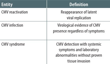
To facilitate interpretation of the presented cases, practical diagnostic categories relevant to critically ill patients are summarised in Table 3.
TABLE 3
Practical interpretation of cytomegalovirus (CMV) detection in critically ill immunocompetent patients
TABLE 4
Clinical manifestations of cytomegalovirus (CMV) disease and diagnostic challenges in critically ill patients
| Clinical manifestation | Typical findings | Diagnostic challenges in ICU patients | Diagnostic considerations |
|---|---|---|---|
| CMV pneumonia | Hypoxia, respiratory failure, diffuse ground-glass opacities, consolidations, tree-in-bud pattern on CT | Radiological findings overlap with ARDS, VAP, pulmonary oedema, and fungal infections | BAL PCR may detect viral shedding rather than invasive disease; BAL viral load thresholds have not been validated for immunocompetent ICU patients. Histopathological confirmation remains the diagnostic gold standard but is rarely feasible in unstable critically ill patients [9, 10]. |
| Gastrointestinal CMV disease | Diarrhoea, abdominal pain, GI bleeding, colitis | Symptoms frequently mimic sepsis-associated ileus, ischaemia, antibiotic-associated diarrhoea, or C. difficile infection | Endoscopic findings are nonspecific. Histopathology with immunohistochemistry remains definitive; PCR positivity alone may overestimate clinically relevant disease [17]. |
| CMV encephalitis | Altered mental status, encephalopathy, seizures, focal neurological deficits | Neurological deterioration may result from trauma, metabolic encephalopathy, sepsis-associated encephalopathy, or drug effects | CSF PCR is highly sensitive; however, interpretation may be difficult in patients with CMV DNAemia or blood contamination. MRI findings are suggestive but not specific. |
| CMV-associated haematological abnormalities | Cytopenias, thrombocytopaenia, coagulopathy, thrombotic events | Haematological abnormalities are common and usually multifactorial in ICU patients | CMV may contribute to immune thrombocytopaenia or coagulopathy; however, causality is often difficult to establish and alternative aetiologies must be excluded [18, 19]. |
These definitions are adapted from published consensus criteria and expert recommendations developed primarily for immunocompromised populations [8, 16] and should be interpreted with caution in critically ill patients without formal immunosuppression.
Importantly, the thresholds for CMV DNAemia used to guide pre-emptive therapy in transplant recipients – typically 100–1000 IU mL–1 – have not been validated in ICU populations and may not reflect the same clinical significance in the context of sepsis-induced immune dysregulation. Moreover, many clinical manifestations traditionally attributed to CMV syndrome – including fever, cytopaenias, liver dysfunction, or respiratory deterioration – are highly prevalent among critically ill patients irrespective of CMV status. Consequently, extrapolation of transplant-based diagnostic frameworks to ICU populations may substantially overestimate the clinical relevance of CMV detection.
The concept of CMV syndrome, originally defined for solid organ transplant recipients, relies on clinical and laboratory criteria that are frequently encountered in critically ill patients for reasons unrelated to CMV – including fever, cytopaenias, and elevated liver enzymes – potentially increasing the risk of overinterpretation when these definitions are extrapolated to the ICU setting. Moreover, histopathological confirmation, which underpins the diagnosis of proven tissue-invasive CMV disease, is rarely achievable in critically ill patients. These limitations underscore the difficulty of applying transplant-based definitions directly to the ICU population, and the definitions presented in Table 3 should be regarded as pragmatic clinical guidance rather than validated diagnostic criteria for this group.
The most important clinical manifestations of CMV disease and associated diagnostic challenges in critically ill patients are summarised in Ta- ble 4 [9, 10, 17–19].
Randomised trials evaluating antiviral prophylaxis demonstrated reductions in CMV reactivation and inflammatory markers but did not show a clear mortality benefit [12, 13], and routine antiviral therapy cannot currently be recommended for all ICU patients with CMV DNAemia. Further evidence is expected from ongoing trials [20].
In clinical practice, delayed availability of virological testing and limited feasibility of histopathological confirmation may complicate therapeutic decision-making in critically ill patients. As a result, clinicians may occasionally face situations in which the potential clinical relevance of viral reactivation must be weighed against the risks of empirical anti-viral therapy despite persistent diagnostic uncertainty. Consequently, interpretation of positive PCR findings requires careful integration with the overall clinical context. For this reason, antiviral treatment decisions in critically ill, immunocompetent patients remain largely individualised and should be interpreted cautiously in the absence of validated diagnostic criteria.
The diagnostic reasoning was particularly relevant in Case 1, in which increasing CMV DNA levels in serial blood samples, together with gastrointestinal symptoms, respiratory failure, and negative microbiological investigations raised suspicion that CMV reactivation may have contributed to the clinical deterioration. Similarly, in Case 2, systematic exclusion of alternative causes made CMV-associated immune thrombocytopaenia a plausible contributing explanation for the clinical presentation.
The second case additionally highlights the possible association between CMV reactivation and severe immune thrombocytopaenic purpura, one of the haematological complications reported in association with CMV infection [21, 22]. Thrombocytopaenia is a common finding in critically ill patients and is usually multifactorial, most frequently associated with sepsis, disseminated intravascular coagulation, drug exposure, or bone marrow suppression. Viral infections, including CMV, may trigger secondary immune thrombocytopaenia.
Standard first-line treatment for immune thrombocytopaenia includes corticosteroids; however, their role in CMV-associated thrombocytopaenia remains controversial because immunosuppressive therapy may potentially enhance viral replication. In addition, CMV infection has been reported to render immune thrombocytopaenia refractory to standard corticosteroid therapy [23]. In the presented patient, corticosteroids were initiated before CMV reactivation had been confirmed, because immune-mediated thrombocytopaenia was considered in the differential diagnosis given the severity of thrombocytopaenia and the need for urgent treatment. Available data regarding CMV-associated thrombocytopaenia in immunocompetent adults remain limited. In the study by Shragai et al. [24], patients with CMV-associated thrombocytopaenia who received anti-CMV therapy demonstrated higher response rates than those managed with steroid-containing regimens alone, suggesting that antiviral therapy may be beneficial in selected patients with severe or refractory CMV-associated thrombocytopaenia. Intravenous immunoglobulin therapy (IVIG) may be considered as adjunctive therapy in selected severe cases of CMV disease, particularly CMV pneumonia, as suggested in recommendations from the European Conference on Infections in Leukaemia (ECIL), although the level of evidence supporting this approach remains limited.
Concurrent administration of ganciclovir, intravenous immunoglobulin, corticosteroids, and platelet transfusions precludes definitive attribution of the clinical response to any single intervention. In the presented patient, the diagnosis was supported primarily by systematic exclusion of alternative causes together with high-level CMV DNAemia. Furthermore, serological evidence supported active CMV reactivation: CMV IgG was positive with IgM negative at presentation, consistent with reactivation rather than primary infection, and a rise in CMV IgG titre was documented on follow-up testing.
Compared with CMV, HHV-6 reactivation in immunocompetent critically ill patients remains less studied. HHV-6 is a neurotropic virus capable of causing severe encephalitis, particularly in transplant recipients and other immunocompromised populations [25, 26]. In immunocompetent adults, reported cases remain rare [14, 27], and interpretation of positive PCR findings is complicated by the possibility of chromosomally integrated HHV-6 (ciHHV-6), in which viral DNA may be persistently detectable despite the absence of active infection. Diagnostic criteria proposed by Bhanushali et al. [28] include clinical symptoms of encephalitis, detection of HHV-6 DNA in cerebrospinal fluid, and exclusion of alternative causes of neurological deterioration.
In Case 3, detection of HHV-6 DNA in cerebrospinal fluid together with marked CSF pleocytosis, refractory hyperthermia, rapidly progressive neurological deterioration, and exclusion of alternative aetiologies supported the suspicion of possible HHV-6-associated encephalitis, although ciHHV-6 could not be excluded due to lack of specialised testing. This substantially limits the certainty of attributing the observed neurological deterioration directly to active HHV-6 infection.
Currently, there are no clear guidelines for the treatment of HHV-6 reactivation in immunocompetent ICU patients. Antiviral therapy with ganciclovir or foscarnet is commonly used in immunocompromised populations [15].
Rather than establishing a definitive causal role of herpesvirus reactivation in critical illness, the presented cases highlight the substantial diagnostic uncertainty associated with interpretation of positive viral PCR findings in ICU patients. The clinical challenge lies not only in identifying viral reactivation, but also in determining whether viral detection reflects clinically meaningful disease, immune dysregulation-associated reactivation, or incidental viral shedding in the context of severe systemic illness.
Conclusions
Critically ill patients following sepsis or major trauma may develop secondary immune dysregulation that can predispose them to latent herpesvirus reactivation.
Distinguishing between asymptomatic viral shedding and clinically relevant viral disease in this population remains challenging due to the nonspecific clinical presentation and the limitations of available diagnostic tools. Careful interpretation of molecular results, including viral load dynamics, together with the clinical context is essential to avoid both underdiagnosis and overtreatment.
Therapeutic decisions in ICU patients are currently largely extrapolated from recommendations developed for immunocompromised populations. Further research is required to better define the clinical significance of herpesvirus reactivation in critically ill patients and to develop evidence-based diagnostic and therapeutic strategies for CMV and HHV-6 disease in immunocompetent ICU populations.