Dear Editor,
This case report describes a 13-year- old girl treated in the intensive care unit for severe methemoglobinemia resulting from intentional poisoning. Methemoglobinemia is characterized by the presence of increased levels of methemoglobin (MetHb), in which the iron within the heme moiety is oxidized to the ferric (Fe3+) state rather than the normal ferrous (Fe2+) state. This alteration impairs hemoglobin’s ability to bind and transport oxygen. Under physiological conditions, the proportion of MetHb does not exceed 2% of total hemoglobin. Clinical manifestations of methemoglobinemia are directly related to MetHb concentration. At levels of 50–70%, patients may develop severe metabolic acidosis, cardiac arrhythmias, respiratory failure, and neurological disturbances, including seizures and coma. MetHb levels exceeding 70% are associated with profound hypoxia, circulatory shock, and death, as reported in the literature [1].
Methemoglobinemia is a rare condition, and its incidence is largely based on isolated case reports, as comprehensive epidemiological data are unavailable. The disorder may be either hereditary or acquired. Congenital forms are uncommon and most frequently result from cytochrome deficiencies or missense mutations affecting globin chains, leading to structurally abnormal hemoglobin. In contrast, acquired methemoglobinemia is more prevalent and is typically associated with exposure to oxidizing agents, including certain medications, contaminated drinking water, food preservatives, and infectious processes – particularly gastrointestinal infections in the pediatric population [2–7]. We confirm that written informed consent for the publication of the article was obtained from the patient’s parents, who are her legal guardians.
A 13-year-old girl was admitted to the Intensive Care Unit (ICU) following initial management in the Emergency Department (ED) for severe methemoglobinemia resulting from intentional sodium hypochlorite ingestion.
The patient was transported to the hospital by an Emergency Medical Team. According to her father, she returned home from a walk holding an almost empty 250 mL bottle of pool-cleaning solution containing sodium hypochlorite. Shortly after entering the house, she developed impaired consciousness with syncope, progressive cyanosis, and profuse vomiting, accompanied by a strong chlorine odor. The father immediately brought her to a nearby primary healthcare facility, from where the ambulance was promptly dispatched. Precise determination of the time of ingestion was not possible. The patient left home at approximately 3:10 p.m., returned shortly after 4:00 p.m., and was registered in the ED at 5:19 p.m. During transport, she was cyanotic and unconscious, with observed seizures, trismus, and recurrent vomiting. Oxygen therapy and intravenous fluids were administered, and upper airway patency was maintained using an oropharyngeal tube.
On admission to the ED, the pa - tient was in a critical condition, presenting with blue-brown discoloration of the skin, unconsciousness, apnea, and absence of a palpable peripheral pulse. Cardiopulmonary resuscitation was immediately initiated. After approximately one minute, spontaneous circulation returned, with a peripheral pulse of 140 beats per minute and restoration of sinus rhythm. Pulse oximetry revealed an oxygen saturation of 55%, and the patient was promptly intubated. Initial laboratory investigations demonstrated severe metabolic acidosis on arterial blood gas analysis (pH 7.11), profound methemoglobinemia (MetHb level 78.1%; reference range 1–1.5%), and a markedly elevated lactate concentration (13.8 mmol L–1; reference range 0.3–0.8 mmol L–1). Due to suspected aspiration, a chest radiograph was obtained, revealing asymmetric lung aeration caused by inadvertent right mainstem bronchial intubation; the position of the endotracheal tube was subsequently corrected. Immediately prior to transfer to the ICU, a telephone consultation with a toxicology specialist was conducted. Administration of methylene blue at a dose of 1–2 mg kg–1 as a 5-minute intravenous infusion was recommended. In the absence of a prompt therapeutic response, repetition of the dose was advised. If treatment remained ineffective, exchange transfusion was recommended as a further therapeutic option.
On admission to the ICU, the patient remained in a critical condition and was unconscious, with a Glasgow Coma Scale score of 8, with preserved responses to deep painful stimuli. Pupils were equal in size and reactive to light. Marked hypersalivation and repeated episodes of serous vomiting were observed, along with generalized muscle rigidity and convulsive activity. Clinical examination revealed severe abnormalities. Hemodynamic assessment demonstrated arterial hypertension (150/90 mmHg) and tachycardia (120 beats min–1). The skin exhibited a gray-brown discoloration. On auscultation, vesicular breath sounds were present bilaterally; however, despite adequate mechanical ventilation and preserved respiratory drive, oxygen saturation measured by pulse oximetry was only 1%.
Laboratory investigations confirmed metabolic acidosis (pH 7.22), significant methemoglobinemia (MetHb 63.2%), hyperkaliemia (6.1 mmol L–1), and elevated lactate concentration (7.2 mmol L–1). Additional findings included leucocytosis (20 × 103 µL–1), with renal and hepatic function parameters within normal ranges (Table 1).
TABLE 1
Renal and liver parameters
According to the initial toxicology consultation obtained in the ED, methylene blue was administered immediately at a dose of 50 mg, followed by a second identical dose after 20 minutes. Due to the lack of an adequate clinical response, therapeutic exchange transfusion was subsequently initiated.
The patient, with a body mass of 40 kg, height of 152 cm, and body mass index of 17.3 kg m–2, was estimated to have a circulating blood volume of approximately 2,800 mL. Owing to rapid clinical deterioration, the exchange transfusion was initially performed using uncrossmatched universal donor blood, followed by group-specific compatible blood once available. Concomitantly, blood products were administered, including fresh frozen plasma (FFP) and platelet concentrate (PC). Multi-electrolyte intravenous fluids were infused, and calcium supplementation was provided as indicated.
Acid–base status (Table 2) and se- rum electrolytes were assessed at re gular intervals, and biochemical para meters were closely monitored throughout the procedure. In total, 16 units of red blood cell concentrate, 6 units of FFP, and 1 unit of PC were transfused, with an overall exchanged blood volume of 4,400 mL.
TABLE 2
Dynamics of changes in blood gas parameters at the beginning and in the first hours of therapy
Following the exchange transfusion, marked cutaneous flushing was observed. Intravenous clemastine (2 mg) and dexamethasone (4 mg) were administered in response. Anal-gosedation was maintained using continuous intravenous infusions of morphine and propofol. Episodes of seizures and generalized muscle rigidity occurred during treatment but resolved following administration of benzodiazepines.
Given the high likelihood of gastrointestinal tract injury, intravenous omeprazole was initiated at a dose of 120 mg over 24 hours. In accordance with the recommendations of the consulting surgeon, endoscopic evaluation was deferred until 24–48 hours after ingestion of the toxic substance.
After approximately two hours, with stabilization of the patient’s clinical condition, the exchange transfusion was completed. Post-procedural arterial blood gas analysis revealed improvement in acid-base status, with a pH of 7.25, a MetHb level of 1.6%, and a lactate concentration of 5.3 mmol L–1. Computed tomography (CT) of the head, chest, and abdominal cavity was subsequently performed and demonstrated no acute abnormalities, in particular no evidence of cerebral edema or gastrointestinal tract perforation. A repeat toxicology consultation did not yield any additional therapeutic recommendations.
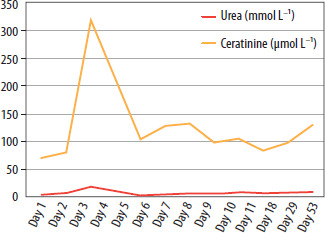
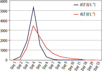
In the hours following completion of the exchange transfusion, a trend toward hypotension was observed. Norepinephrine was initiated at a dose of 0.1 µg kg–1 min–1, resulting in an adequate hemodynamic response. Owing to coagulation abnormalities, the patient required repeated transfusions of FFP. Renal and hepatic function parameters remained within normal ranges during the first 24 hours; however, over subsequent days the patient developed acute liver failure, with markedly elevated aminotransferase levels (aspartate aminotransferase test [AST] 1,924 U L–1; alanine aminotransferase test [ALT] 1,217 U L–1), as well as acute kidney injury necessitating initiation of renal replacement therapy (Table 1). Peak urea and creatinine concentrations reached 18.5 mmol L–1 and 316.6 µmol L–1, respectively. Oliguria was also observed, with urine output on the second day of hospitalization measuring 0.7 mL kg–1 h–1 (730 mL over 24 hours). Continuous veno-venous hemodiafiltration was performed for 11 days. Subsequently, due to persistently elevated renal function parameters despite restoration of adequate diuresis, intermittent hemodialysis was initiated (Figure 1). Episodes of arterial hypertension were noted during the clinical course, and antihypertensive therapy was introduced. Hepatic enzyme levels gradually normalized within several days (Figure 2).
In accordance with surgical recommendations, endoscopic evaluation of the gastrointestinal tract was performed 24 hours after ingestion. The examination confirmed extensive injury, revealing diffuse esophageal edema along its entire length, mucosal erythema, and severe gastric involvement characterized by extensive mucosal burns, marked edema, deep ulcerations, and contact bleeding. Given the high risk of gastrointestinal perforation, enteral feeding was initially contraindicated. Total parenteral nutrition was therefore initiated, and intravenous proton pump inhibitor therapy was continued. Ornithine was administered at a dose of 10 g per day. In the subsequent days, follow-up endoscopy was performed, during which a feeding tube was placed, and enteral nutrition with a peptide-based formula was initiated with gradual escalation of volume.
Prior to the decision to discontinue pharmacological sedation and awaken the patient, a control CT scan of the head was obtained, which demonstrated no acute abnormalities. Hemo-dynamic status gradually stabilized; however, norepinephrine support was required until the seventh day of ICU hospitalization.
With gradual improvement in the patient’s condition, sedative medications were progressively tapered, and on the 11th day of ICU hospitalization, the patient was extubated and transitioned to spontaneous breathing, initially supported by supplemental oxygen. Neurologically, she exhibited no significant deficits and was fully oriented and alert. However, she was reluctant to speak and displayed low mood, prompting engagement of psychological support. In the subsequent days, communication remained limited, not only with medical staff but also with her parents. The attending psychologist recommended urgent psychiatric consultation; however, a comprehensive psychiatric assessment could not be performed due to minimal verbal interaction. During the psychiatric evaluation, the patient responded only with head nods and did not engage in conversation. The consulting psychiatrist advised a follow-up psychiatric assessment once the patient’s somatic condition further improved and recommended ongoing psychological support for both the patient and her parents.
After three weeks of hospitalization, the patient was transferred to the Gastroenterology Department for continued care. Hemodialysis was maintained every 2–3 days for 17 days, according to the patient’s clinical status. Enteral and parenteral nutrition were administered based on tolerance. During attempts to gradually reintroduce oral feeding over the course of more than one month, the patient experienced recurrent symptoms, including abdominal pain, nausea, and vomiting. Eventually, she achieved full tolerance of an oral diet.
Subsequently, the patient underwent a full psychiatric evaluation, which led to a diagnosis of adjustment disorder, with a recommendation for continued monitoring for potential depressive disorders. After a total hospitalization of two months, she was discharged home in a stable condition, with full tolerance of an oral diet and normalization of renal function. Outpatient psychiatric and psychological follow-up was arranged to support ongoing recovery.
The most commonly reported causes of methemoglobinemia resulting from poisoning include chemotherapeutic agents [7], local anes thetics used during endoscopic procedures [2, 3, 6], nitrogen-containing agricultural compounds [4, 5], and food oxidants [8]. To date, no cases of intentional sodium hypochlorite ingestion leading to methemoglobinemia and requiring advanced intensive care interventions have been reported. In previously described cases [2–8], management with supplemental oxygen therapy, and in some instances methylene blue administration, was sufficient to achieve clinical improvement.
In the present case, it was difficult to determine the exact dose of so dium hypochlorite ingested, as the estimated volume of fluid consumed was approximately 250 mL. Nevertheless, laboratory findings revealed severe methemoglobinemia, indicating substantial absorption of the toxic substance from the gastrointestinal tract. Therapy in this case was guided by both clinical symptoms and the overall MetHb level. According to the literature, asymptomatic patients with MetHb levels below 20% typically do not require specific interventions beyond avoidance of oxidizing agents. Treatment is generally indicated at MetHb levels of 20% in symptomatic patients and 30% in asymptomatic patients [8]. In this patient, severe clinical manifestations were present, including impaired consciousness and respiratory and circulatory compromise, accompanied by markedly elevated MetHb levels. Additionally, the response to methylene blue was minimal, prompting the decision to perform an exchange transfusion as a definitive measure to remove MetHb from the vascular compartment.
An additional therapeutic challenge in this case was the limited availability of monitoring techniques for accurately assessing the patient’s oxygenation status. Conventional pulse oximetry is unreliable in cases of methemoglobinemia, and standard blood gas analysis only measures dissolved oxygen in plasma rather than the oxygen content bound to hemoglobin. Standard pulse oximeters estimate oxygen saturation using two wavelengths of light (red and infrared), assuming that only oxyhemoglobin and deoxyhemoglobin are present.
MetHb absorbs both wavelengths equally, leading to a constant, falsely elevated oxygen saturation reading of approximately 80–88%, regardless of the true oxygenation status. Co-oximetry represents the method of choice in such situations, provided that the equipment is available in the ICU. Co-oximeters utilize at least four wavelengths of light to differentiate and quantify multiple hemoglobin species, including oxyhemoglobin, deoxyhemoglobin, carboxyhemoglobin, and MetHb, allowing for accurate assessment of oxygen-carrying capacity [9]. Repeated and thorough clinical assessment, combined with rapid implementation of targeted therapy, was crucial in stabilizing the patient’s condition and preventing permanent organ damage.
The importance of a multidisciplinary approach should be emphasized, as management involved toxicology and nephrology consultations, alongside timely psychological and psychiatric support. Suicide attempts are an increasingly frequent cause of pediatric intensive care admissions. In cases of poisoning, there is a high risk of long-term organ complications. These considerations highlight the importance not only of prompt pre-hospital and hospital management but also of preventive strategies, including psychoeducation programs in schools to address mental health and reduce the risk of self-harm.
In conclusion, suicide attempts are an increasingly frequent cause of pediatric intensive care admissions, and cases involving poisoning carry a high risk of long-term organ complications. The case presented here represented a significant therapeutic challenge, not only due to the need for intensive, dynamic, and multi-modal management, but also because of limitations in monitoring capabilities.
The primary therapeutic goal was to prevent hypoxia, which could have led to catastrophic complications affecting the nervous system, kidneys, and liver, with potential long-term consequences. Conventional pulse oximetry is unreliable in cases of methemoglobinemia, and standard blood gas analysis only measures oxygen dissolved in plasma rather than oxygen bound to hemoglobin. Through repeated and thorough clinical assessments, careful interpretation of laboratory results, and intensive multi-disciplinary therapy, the patient’s neurological outcome was ultimately normal.
Overall, it can be concluded that, as a result of comprehensive, targeted treatment, the patient avoided clinically significant hypoxia and permanent organ damage, and the observed transient organ dysfunction was most likely attributable to direct toxic effects of sodium hypochlorite rather than secondary hypoxic injury.