Introduction
The management of neuromuscular blocking agents (NMBAs) during anaesthesia remains a critical component of perioperative patient safety. Residual neuromuscular blockade (RNMB) is associated with adverse outcomes such as respiratory complications, delayed extubation, and increased postoperative morbidity [1, 2]. Quantitative neuromuscular monitoring is therefore widely recommended to ensure adequate onset and recovery of neuromuscular function prior to intubation and recovery of consciousness [3–5]. A recent consensus statement emphasises that objective monitoring should be used whenever NMBAs are administered, and that a train-of-four (TOF) count £ 3 is commonly used as threshold for ideal intubation conditions, and a TOF ratio ≥ 0.90 is required to reliably exclude RNMB [4, 6].
Traditionally, neuromuscular blockade has been assessed using mechanomyography (MMG) – the measurement of muscle force in response to peripheral nerve stimulation – which has been considered the “gold standard” in research settings [6, 7]. However, because MMG is cumbersome for routine clinical use, acceleromyography (AMG) historically became the most widely used clinical standard. More recently, electromyography (EMG), which monitors the evoked electrical response of the muscle following nerve stimulation [8], has increasingly been advocated as a practical alternative to AMG. EMG offers precise, quantitative measurement of ratios and enables accurate assessment of neuromuscular recovery without the mechanical limitations asso-ciated with movement-based monitors [1, 4].
Kinemyography (KMG) represents another commercially available and clinically utilised quantitative monitoring technique. KMG measures the mechanical deformation or movement of a muscle in response to nerve stimulation, rather than the electrical signal measured by EMG or the force measured by MMG [9]. Because KMG is relatively easy to apply and uses simple movement-sensing elements, it has attracted interest in clinical neuromuscular monitoring. For example, one paediatric study showed excellent agreement between KMG and EMG for cisatracu-rium onset and recovery times [10]. However, stu-dies in adults have shown that, although KMG demonstrates acceptable precision, it tends to overestimate TOF ratios during the recovery phase compared with EMG, with wide limits of agreement that may limit its clinical interchangeability for excluding RNMB [8, 9]. To our knowledge, no study has directly evaluated the clinical impact of the wide limits of agreement between these two monitoring methods during recovery, and importantly not during the onset phase. Consequently, there remains a gap in knowledge regarding the clinical implications of KMG TOF ratio overestimation for intubation and extubation timing.
Given these findings, a direct clinical comparison of KMG versus EMG in adult patients under neuromuscular blockade is warranted. A better understanding of the comparative clinical performance of these two modalities may inform clinical practice guidelines and influence the choice of monitoring technology in the operating theatre. Therefore, we conducted this study to evaluate neuromuscular monitoring using KMG and EMG during both the onset and recovery phases of non-depolarising neuromuscular blockade.
We hypothesised that KMG systematically overestimates TOF ratios compared to EMG. The primary objective of the study was to compare the onset and recovery times of rocuronium-induced neuromuscular blockade as measured by EMG and KMG. The secondary objective was to compare the T1% parameter between patients who received sugammadex for reversal of neuromuscular block and those in whom neuromuscular function recovery occurred spontaneously, meaning without admi-nistering sugammadex or another antidote.
Methods
We conducted a prospective observational study in adult patients undergoing general anaesthesia with administration of rocuronium. The study took place between 11 December 2023 and 25 July 2024 in Ústí nad Labem Czech Republic in Masaryk Hospital Anaesthesia Department. The study was approved by the Ethics Committee of Masaryk Hospital Usti nad Labem (reference number 316/1) and was registered at ClinicalTrials.gov (registration number NCT05992090). The study was conducted in accordance with the Declaration of Helsinki and good clinical practice. All patients agreed to be included in the clinical trial and signed an informed consent form.
Study design
After signing an informed consent form, patients were transported to the operating theatre. Standard monitoring of vital signs, depth of anaesthesia, and depth of neuromuscular blockade was applied in all cases.
For quantitative neuromuscular monitoring, we compared two different plug-in sensor modules (KMG and EMG) connected to the same anaesthesia monitoring system (Carescape One, model type 2087075-300, manufactured in 2023, country of origin Finland, software version 3, software build 3.2.772, GE Healthcare, Madison, Wisconsin, USA) with a Carescape Canvas Smart Display (model type 587060, manufactured in 2023, country of origin Finland, software version 3.3.377; GE Healthcare, Madison, Wisconsin, USA). Both the KMG sensor (model type E-NMT-01, manufactured in 2018, country of origin Finland, GE Healthcare, Madison, Wisconsin, USA) and the EMG sensor (model type E-NMT-01, manufactured in 2018, country of origin Finland, GE Healthcare, Madison, Wisconsin, USA) were used simultaneously. The KMG sensor was placed on the right hand and the EMG sensor on the left hand during odd study weeks, with the configuration reversed during even weeks.
Simultaneous KMG and EMG monitoring was performed using two independent monitors. EMG was used as the reference standard to guide clinical decision-making. The attending anaesthesiologist was blinded to KMG measurements, had no visual access to the KMG monitor, and was not informed of KMG values throughout the procedure.
EMG neuromuscular monitoring was performed by stimulating the ulnar nerve at the wrist and recording from the adductor pollicis muscle. Two stimulation electrodes were placed over the course of the ulnar nerve at the volar aspect of the wrist, with the distal (cathode) electrode positioned 1–2 cm proximal to the wrist crease and the proximal (anode) electrode placed 2–3 cm proximally along the nerve. The recording electrode was positioned over the belly of the adductor pollicis muscle, with the reference electrode placed on the proximal phalanx of the thumb. A ground electrode was placed on the distal forearm between the stimulation and recording sites.
KMG neuromuscular monitoring was performed by stimulating the ulnar nerve at the wrist and measuring thumb movement using a piezoelectric sensor. Two stimulation electrodes were placed over the course of the ulnar nerve at the volar aspect of the wrist, with the distal (cathode) electrode positioned 1–2 cm proximal to the wrist crease and the proximal (anode) electrode placed 2–3 cm proximally along the nerve. The KMG sensor was secured to the volar surface of the thumb to detect contraction of the adductor pollicis muscle. The hand and fingers were positioned according to the manufacturer’s recommendations to allow unrestricted thumb movement during stimulation.
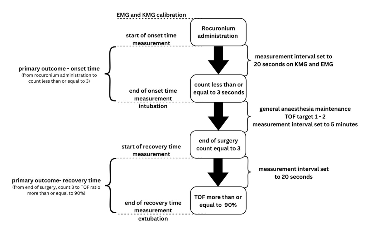
General anaesthesia was induced with a bolus of propofol at a dose of 2 mg kg–1 and sufentanil at 0.2 µg kg–1. Both KMG and EMG monitors were then calibrated using the manufacturers’ integrated calibration procedures. Neuromuscular monitoring was subsequently initiated with measurements obtained at 20-second intervals.
Following calibration, rocuronium was admini-stered at a dose of 0.6 mg kg–1 ideal body weight.
Tracheal intubation was performed at a TOF count less than or equal to 3. While recent guidelines advocate for deeper blockade prior to intubation, this threshold was strictly dictated by our local standard operating procedures at the time of study design and remains in line with historical consensus for acceptable intubating conditions [5]. The onset time was measured.
During maintenance of anaesthesia, additional doses of rocuronium were administered as needed to maintain a TOF count of 1–2. Because our study endpoints focused exclusively on the onset and recovery phases, this moderate block maintenance strategy, while suboptimal for procedures such as robotic surgery, did not influence the primary outcomes. Anaesthesia maintenance was performed using sevoflurane and sufentanil, with no protocol-based dosing. Sufentanil was administered to maintain the Surgical Pleth Index between 30 and 50.
At the end of the surgical procedure, emergence from anaesthesia was initiated. The recovery time was measured. During this phase, the neuromuscular monitoring interval remained set at 20 seconds.
Administration of sugammadex at a dose of 2 mg kg–1 or neostigmine with atropine (0.05 mg kg–1 neostigmine with 0.5 mg atropine) was left to the discretion of the attending anaesthesiologist, without a predefined decision protocol. Extubation was performed once a TOF ratio ≥ 0.90 was achieved.
Following extubation, all patients were transferred to the post-anaesthesia care unit, marking the end of their participation in the study. A flowchart of the data collection is presented in Figure 1.
Study population
Inclusion criteria were adult patients (≥ 18 years) undergoing general anaesthesia with administration of rocuronium. Exclusion criteria included the presence of a pre-existing neuromuscular disorder (such as myasthenia gravis, Lambert-Eaton mya-sthenic syndrome, polymyositis, dermatomyositis, Duchenne and Becker muscular dystrophy, Pompe disease, and mitochondrial myopathies), known allergy to adhesive electrode materials, emergency case, BMI > 40, difficult access to both hands during surgery, and refusal to participate.
Data collection
Neuromuscular monitoring data from both KMG and EMG systems were recorded simultaneously at predefined time points by a dedicated study nurse. Data were collected throughout induction, maintenance, and recovery phases of anaesthesia, including onset and recovery of neuromuscular blockade.
Outcome measures
The primary outcome measures were onset time and recovery time of neuromuscular blockade. Onset time was defined as the time from rocuronium administration to achievement of a TOF count ≤ 3. Recovery time was defined as the interval from a first TOF count of 3 read at the end of the surgical procedure to attainment of a TOF ratio ≥ 0.90.
The secondary outcome measure was T1% measured at the time of extubation. T1% was defined as the ratio of the amplitude of the first twitch in each twitch series to the amplitude of the first twitch recorded during the calibration procedure. The clinical rationale for exploring this normalised T1% parameter is to assess the absolute recovery of muscle strength, which may remain depressed even when the TOF ratio (which primarily measures fade) has successfully recovered to over 0.90.
Statistical analysis
A sample size of 94 subjects was calculated based on pilot data to identify a 20% difference in the primary outcome between the two groups with 90% power with a cut-off for statistical significance of P = 0.05. A group size was set to 130 participants with respect to potential loss to analysis. The mean values ± standard deviation (SD) or percentages were calculated as necessary. The normality of the data was evaluated according to the Kolmogorov-Smirnov test. Differences in categorical variables were standardly evaluated using the c2 test and Pearson coefficient. Comparisons of two continuous variables were calculated using t-tests for parametric variables or the Mann-Whitney U test for non-parametric variables. All calculations were performed in an open-source R environment and plotted using the ggplot2 library (v4.1.2, R Core Team [2021]; R is a language and environment for statistical computing from the R Foundation for Statistical Computing, Vienna, Austria, https://www.R-project.org/).
This study was conducted and reported in accordance with the STROBE recommendations for observational research, and the completed checklist is provided as supplementary material.
Results
We enrolled 137 patients in total, including 73 males (53%) and 64 females (47%). The mean body mass index was 28.2 ± 7.4 kg m–2, and the mean age was 54 years. Demographic and baseline characteristics of the study population are presented in Table 1.
TABLE 1
Demography of studied population
Regarding the management of neuromuscular blockade recovery, 52 patients (38%) received sugammadex and 85 (62%) patients recovered spontaneously without any pharmacological reversal. There were no patients who received neostigmine with atropine as reversal.
The time required to reach a TOF count ≤ 3 following rocuronium administration (onset time) was significantly longer when assessed by EMG compared with KMG (158 ± 74 s vs. 118 ± 68 s). The mean paired difference was 40 seconds (95% CI 22.9–57.1; paired t-test, P < 0.001), favouring earlier detection by KMG.
Recovery time was also longer with EMG (27 ± 22 min) compared with KMG (24 ± 21 min). The mean difference was 3 minutes (95% CI: –2.2 to 8.1; paired t-test, P = 0.242), indicating no statistically significant difference between methods.
The standardised effect size (Cohen’s d) for onset time was 0.56 (95% CI: 0.32–0.81), consistent with a moderate effect, whereas for recovery time it was 0.09, indicating a negligible effect. Detailed results for the primary outcomes are presented in Table 2.
TABLE 2
Results for primary outcomes
In 95 cases (69%), EMG detected a TOF count ≤ 3 later than KMG; in 25 cases (18%), both methods detected it simultaneously; and in 17 cases (12%), KMG detected a TOF count ≤ 3 later than EMG.
The coefficient of variation during onset was 0.47 for EMG and 0.58 for KMG, indicating mode-rate relative dispersion. During recovery, relative variability was higher, with coefficients of variation of 0.81 for EMG and 0.87 for KMG. Across both phases, KMG demonstrated slightly higher relative variability.
RNMB (defined as TOF ratio < 0.90 at extubation) was observed more frequently in patients who did not receive sugammadex compared with those who did (3 vs. 0 cases; Fisher’s exact test, P = 0.031). Mean T1% at extubation was higher in patients receiving sugammadex compared with spontaneous recovery (77 ± 18% vs. 70 ± 21%; independent samples t-test, P = 0.074), as detailed in Table 3.
TABLE 3
Comparison of mean T1% parameter and residual neuromuscular block in patients who received sugammadex with those who did not
| Factor | Sugammadex not administered | Sugammadex administered | P-value |
|---|---|---|---|
| n (%) | 85 (62) | 52 (38) | |
| T1% | 70 ± 21 | 77 ± 18 | 0.074 |
| RNMB, n (%) | 3 (4) | 0 (0) | 0.031 |
In an exploratory post hoc subgroup analysis of patients who did not receive sugammadex, recovery time showed a non-significant trend toward longer duration when assessed by EMG compared with KMG (42 ± 19 min vs. 37 ± 18 min; paired t-test, P = 0.183). This analysis was conducted to explore whether reversal strategy influenced agreement between monitoring modalities and should be interpreted as hypothesis-generating.
Discussion
Our primary findings demonstrate a clinically meaningful difference between KMG and EMG moni-toring during both onset and recovery of neuro-muscular blockade. EMG measured significantly longer onset times compared with KMG (mean difference 40 s), while recovery times showed a similar directional pattern, although without statistical significance. KMG detected a TOF count ≤ 3 earlier in 69% of cases, supporting the conclusion that the two modalities are not interchangeable during onset of neuromuscular blockade. These results highlight the importance of careful consideration when selecting neuromuscular monitoring technology in clinical practice.
The clinical relevance of accurate neuromuscular monitoring is well established [11]. RNMB is associated with an increased risk of postoperative pulmonary complications, impaired upper airway function, aspiration, and hypoxic events [11]. Current consensus guidelines therefore recommend objective quantitative monitoring and a TOF count of 0 for ideal intubation conditions, and a TOF ratio ≥ 0.90 prior to extubation [4, 5, 11, 12]. Because our results demonstrate a delay in EMG-defined onset compared with KMG, reliance on KMG alone for determining optimal intubation timing may risk inadequate suppression of laryngeal reflexes – potentially leading to coughing, laryngospasm, vocal cord injury, and airway trauma [6, 13, 14].
The discrepancy between EMG and KMG during recovery phase has been previously described [8, 9]. However, these studies focused primarily on the mathematical agreement of the modalities only during recovery. To our knowledge, the direct clinical impact of this discrepancy has been completely lacking in the literature. Our study directly addresses this gap. By evaluating the actual time differences to reach clinical thresholds, our study demonstrates how the previously described limits of agreement translate into real-world clinical consequences regarding the safe timing of extubation.
During deep neuromuscular block, small mechanical responses may persist despite near-complete transmission failure at the neuromuscular junction [4]. This can lead to micromovements or desynchronised movements and therefore syste-matic overestimation of TOF and accelerated interpretation of recovery [9]. Our finding that EMG indicated deeper block during onset in 95 out of 137 cases supports this physiological principle.
The clinical implications extend beyond the induction phase and intubation timing. Misinterpretation of recovery can lead to delayed recognition of persistent neuromuscular impairment and wrong extubation timing. Liu et al. [14] demonstrated that RNMB remains prevalent in modern practice despite the availability of reversal agents, and it may contribute to postoperative pulmonary complications. Although all patients reached a TOF ratio of over 0.90 on EMG prior to extubation, 3 patients in the spontaneous recovery group subsequently exhibited clinical signs of RNMB (e.g. inadequate head lift, paradoxical orofacial muscle activity). This is consistent with evidence that a TOF ratio of more than 0.90 does not universally exclude residual weakness because up to 2–5% of patients may remain symptomatic at this threshold [4]. In these cases, the clinical diagnosis of RNMB was confirmed by the immediate resolution of symptoms following sugammadex administration. The higher-than-expected incidence of these cases in our cohort probably reflects the increased clinical vigilance and sensitivity of the anaesthesiologists towards neuromuscular recovery resulting from their involvement in this prospective monitoring study. Although recovery-time differences between monitoring modalities did not reach statistical significance, the trend toward faster KMG-measured recovery suggests that clinicians may overestimate readiness for safe extubation when relying solely on KMG.
Interestingly, T1% values were higher within the subgroup of patients receiving sugammadex, but not significantly. This finding may partly explain the lower postoperative pulmonary complication rates reported in patients receiving sugammadex in previous studies [14]. In addition, our results explore a novel EMG-derived parameter, T1%. By assessing the absolute recovery of muscle strength relative to baseline, T1% adds valuable information to the standard TOF ratio, which reflects only the degree of fade. Consequently, T1% may represent a promising metric that could potentially provide an additional safety threshold in EMG-based neuro-muscular monitoring, helping to identify patients with generalised residual weakness despite a recovered TOF ratio. To our best knowledge, this study is the first to systematically report on this parameter in a clinical setting. Although investigated as an exploratory outcome, we believe these initial data will serve as a valuable foundation for future studies to establish its definitive clinical role. However, further prospective validation is required before its widespread clinical adoption.
An important strength of our study is the simultaneous bilateral monitoring using an identical device and software, reducing instrument-related variability expressed in clinical end points. Nevertheless, several limitations should be acknowledged. First, this was a single-centre study with recruitment limited to elective surgical cases, so results may not generalise to emergency or critically ill populations. Second, while both sensors were alternated weekly between hands to reduce placement bias, subtle differences in electrode contact or limb position may still affect readings. Third, although sample size calculations ensured adequate power for primary outcomes, subgroup analyses, particularly those involving reversal agents, were exploratory and underpowered for definitive conclusions. Lastly, in our study, the decision to administer sugammadex was left to the clinician’s discretion and was not protocolised, introducing potential confounding and selection bias. Patients perceived to be at higher risk may have been more likely to receive reversal, which limits causal interpretation of subgroup findings.
Although our findings support the more conservative performance profile of EMG during both phases of neuromuscular block (onset and recovery), it is important to recognise workflow realities. KMG devices are easier to apply and may be more widely available, particularly in centres without dedicated quantitative monitoring programs. Therefore, the question that remains is not whether EMG is superior, but how to address barriers to its broader implementation, including cost, user-experience, and lack of mandatory standards. Bridging these gaps may have a greater impact on patient safety than modality refinement alone.
Regarding patient safety, our study highlights that clinicians should maintain heightened awareness of RNMB risk and consider routine sugammadex reversal mainly in cases where KMG is used. Future studies should investigate the role of the T1% parameter in managing neuromuscular blockade because, to our best knowledge, there are no data on the use of T1% parameter, which is available only on an EMG sensor.
Conclusions
Our study demonstrates clinically relevant disagreement between KMG and EMG in the assessment of neuromuscular block during onset and recovery in adults. These findings indicate that KMG cannot reliably replace EMG and suggest that EMG-based monitoring may represent a safer approach for timing of intubation and extubation.